Resident: Adam J. Bottrill
Date: 26FEB10
Region: Providence
Article title: Herpetic gingivostomatitis and teething difficulty in infants
Author(s): King, David et. al.
Journal: Pediatric Dentistry
Page #s: pp. 82-85
Year: March/April 1992
Major topic: Primary herpetic gingivostomatitis (PHG)
Minor topic(s): Teething
Type of Article: Retrospective investigation
Main Purpose: The aim of this study was to determine whether PHG may be responsible for signs and symptoms commonly attributed to teething in infants.
Overview of method of research: Patient study and comparison. 20 infants with parental diagnosis of “teething difficulty.” 20 infants serving as controls.
Key points in the article discussion:
I. Oral swab obtained from the infants and processed to determine presence of HSV.
A. Temp and oral status recorded.
B. 9 subjects positive for HSV
C. 7 of 9 had temperature
D. All 9 had varying degrees of visible oral infection
E. Of the remaining 11, 5 had temperatures and none had visible infeciton (otitis media, vericella).
F. The control group was negative for HSV, temp AND visible infection.
II. Results: HSV should be included in differential Dx of patients with CC of “teething difficulty”
A. Finding of HSV “highly significant” in the DDX of teething difficulty.
B. 80-90% of adults have serum AB’s to HSV. Many don’t remember having it. This could be due to undiagnosed infant PHG.
C. Infants with teething difficulty should be more closely analyzed for PHG.
Assessment of article: Short and Sweet. Negative on the Shenanigans.
Thursday, February 25, 2010
Revised pediatric HIV classification system
Resident: J. Hencler
Date: 02/26/2010
Article title: Revised pediatric HIV classification system
Author(s): AAPD
Journal: Pediatric Dentistry-18:2,1996
Major topic: HIV
Type of Article: Revision
Key points in the article discussion:
In 1994, a new classification system for pediatric HIV replaced the 1987 system (see tables)
Category N (not symptomatic): Includes children who have no signs or symptoms considered to be the result of HIV infection or who have only one of the conditions listed in Category A.
Category A (mildly symptomatic): Children with two or more of the conditions listed below but none of the conditions listed in Categories B and C. (see list)
Category B (moderately symptomatic): Children who have symptomatic conditions other than those listed for Category A or C that are attributed to HIV infection. Examples of conditions in clinical Category B include but are not limited to: (see list)
Category C (severely symptomatic): Serious bacterial infections, multiple or recurrent (i.e., any combination of at least two culture-confirmed infections within a 2-year period), of the following types: septicemia, pneumonia, meningitis, bone or joint infection, or abscess of an internal organ or body cavity (excluding otitis media, superficial skin or mucosal abscesses, and indwelling catheter-related infections). (see list)
Summary of conclusions: New pediatric HIV classification system is simpler. It would be good to have a reference manual if needed.
Assessment of article: This revision of the pediatric HIV classification system is good to be aware of because many signs and symptoms of HIV infection can manifest in the oral cavity. This classification is also pertinent to us as pediatric dentists when consulting with a Pediatrician about a child’s HIV status and severity.
Pediatric AIDs
Department of Pediatric Dentistry
Lutheran Medical Center
Name: Craig Elice Date: February 26, 2010
Article title: Pediatric Aids
Author(s): Chadwick EG, Yogev R.
Journal: Dental Clinics of North America
Volume (number): 42(4) 969-979
Month, Year: August 1995
Major topic: summary of Pediatric Aids
Type of Article: Review of Literature
Main Purpose: The article provided an overview of the HIV and AIDs.
1 million of 13 million infected persons are children with 80% in sub-saharan populaton. The prenatal transmission rate is 20-30%. Approximately 89% of infected children less than 13 years of age were infected by vertical transmission which is increasing while less than 10% are infected through contaminated blood products. African Americans represent 15% of the childhood population yet represent 50 % pf the pediatric AIDs cases. Adolescents between 13 and 19 represent the fastest growing segment of Aids cases. Risk factors vary by gender in adolescents. 45% of males with AIDs are infected by contaminated blood or blood products while females are infected by heterosexual contact. Transmission: In pediatric populations, vertical transmission is most common with 12-20% transmission rates in US and Europe and igher transmission rates in Africa and Haiti. This vertical transmission can occur intrauterine, during delivery, or less often via breast feeding. Transfusion s of infected blood account for 9% of pediatric AIDs and it appears to be diminishing as a result of HIV antibody screening. HIV is rarely isolated from saliva and does not appear to be a real danger of transmission. Pathogenesis: HIV binds to cells with CD$ molecules (T4 lymphocytes) which migrate to lymph nodes where they become activated and proliferate. Patients develop an immune response which suppresses HIV levels yet causes a deterioration of the immune response. This suppression phase is the clinical “Latency phase”. Eventually the immune system deterioration frees the virus to recirculate through many body tissues Clinical Manifestations: The CDC pediatric guidelines include status of disease: mild to moderate to severe signs and symptoms as well as the immune classification based of absolute T-helper cells count or the Percent of CD4 cells. Recurrent bacterial infections, chonic parotid swelling, LIP, and early onsey of progressive neurological deterioration are characteristic of pediatric AIDs. Infections: The most common serious infections affecting greater than50% of infected children are bacteremia, sepsis, and pneumonia. While adults experience reactivation of latent infections like viruses, most pediatric infections are primary including pneumocystis carinii pneumonia (PCP). Oral candidiasis is the most common fungal disease in HIV infected children. Viral infections like JSV, VZV, and CMV are common. Systemic: CNS involvement in vertically infected children is approximately 20-50% and presents as progressive encephalopathy with loss or stagnation of milestones, cognitive deterioration and stunted brain growth. Recurrent respiratory like otitis and sinusitis are common. LIP affects 1/3 of children presenting as lymphoid hyperplasia of the bronchia tubules causing blockage of the alveolar/ capillary interface. PCP is the most common opportunistic infection in pediatric AIDs Cardiac involvement in patients with HIV infection has been thoroughly studied. GI involvement is common with symptoms of chronic and recurrent diarrhea with malabsorption abdominal pain, dysphagia, and failure to thrive (FTT). Chronic liver inflammation is common. Renal disease is an uncommon finding. Skin diseases although present in healthy children, are common in HIV infected children. Recurrent and chronic primary infections with HSV, Zoster, and other viruses are common as well as fungal diseases as well as hyperkeratosis. Malignant diseases are rare in pediatric AIDs. Diagnosis is confirmed using immunoassay for IgG antibody to HIV after 18 months of age, while diagnosis is made by virus culture in infants. Management: Focus is placed on nutritional status of patients and prevention of PCP which is difficult as PCP sometimes precedes the diagnosis of AIDs. Guidelines from the CDC for prophylaxis target infants born to infected mothers or shown to be infected by HIV. Therapy includes use of Zidouridine as the first drug of choice in children who have a reduced CD4 count and symptoms of AIDs. Combinations of other meds like Didanosine and others are being investigated. Prognosis is poor when the patient is symptomatic during the first year of life. Opportunistic infections, encephalopathy also carry a poor prognosis.
Key points/Summary: Describes the pathophysiology of pediatric AIDs.
Assessment of article: Good article but may need an update.
Herpes virus Infections
Department of Pediatric Dentistry
Lutheran Medical Center
Name: Craig Elice Date: February 26, 2010
Article title: Herpes virus Infections
Author(s): Greenberg MS.
Journal: Dental Clinics of North America
Volume (number): 40:2 pages 359-368
Month, Year: April 1996
Major topic: summary of herpes viruses
Type of Article: Review of Literature
Main Purpose: The article provided an overview of the herpes viruses that infect human beings.
Of the 80 known viruses, only 7 Herpes viruses infect humans: HSV I and 2, Varicella Zoster (VSV), Cytomegalovirus (CMV), Epstein- Barr (EBV), HHV 6 and HHV 7. Characteristics that they share include: All viruses cause a primary infection upon first patient exposure and become latent within nuclei of certain body cells for the life of the patient. HSV I, 2, and VZV become dormant in sensory nerve ganglia, CMV in lymphocytes and salivary glands, EB in B-lymphocytes and salivary gland tissues, HHV 6 and 7 in CD4 lymphocytes. They recur as localized symptomatic or asymptomatic recurrent infection., Are transmitted by direct contact via saliva or genital secretions. The EBV can transform cells into maligniancy like nasopharyngeal carcinoma Burkitts lymphomaor B cell lymphoma. HSV 1 and 2: HSV 1 is primarily transmitted by saliva and causes most oral, pharyngeal, eye and CNS infections, while HSV 2 is transmitted by genital secretions and casues genital and anal infections. The incidence of primary infections for HSV 1 is after 6 months of age and peakes between 2-3 years of age. Primary infections are often subclinical but can be characterized by fever, chills, nausea, and lymphadenopathy and followed in 1-2 days by vesicles and ulcers of oral tissues and gingivitis. 20-40% of patients develop recurrent infections which are activated by local trauma or systemic fever or menstrual cycle. Diagnosis is made clinically but viral culture and cytology smears and serology can confirm the diagnosis. CMV is mostly asymptomatic in humans except as a congenital infection called Cytomegalovirus inclusion disease. It is transmitted via birth canal secretions, breast milk, saliva in young children, contaminated blood transfusions, and sexual contact. It is a cause of disease in immunocompromised patients like transplant and AIDs patients. Diagnosis is made by histopathologic studies sowing enlarged owl eye cells. VSV: In 0.3-0.5% of the infected patients, the virus becomes activated presenting as herpes zoster. C-3, T-5, L-1 and 2, are the nerves tracts most commonly infected, but can infect the trigeminal. The symptoms include pain, tenderness and paresthsia along the course of the infected nerve which often precedes vesicles. These vesicles along the nerve tract are a diagnostic sign. In immunocompromised patients, the disease can be life threatening. Acyclovir is effective in shortening the duration and accelerates healing. EBV: Although mild in children, in adolescents and young adults, it can lead to infective mononucleosis. Mono has an incubation period of up to 8 weeks and presents as fever, malaise, pharyngitis, and lymphadenopathy. Diagnosis is based on clinical signs and detection of large lymphocytes in the peripheral blood. Oral lesions include hairy leukoplakia HHV 6: The virus infects CD$ lymphocytes and other white blood cells causing roseola which is self limiting. HHV 7: Usually transmitted by saliva and is found in 80% of adults and 70% if children. NO clinical disorder have been associated with it.
Key points/Summary: Describes 7 human herpes viruses.
Assessment of article: Good article but may need an update.
Oral manifestations of HIV in kids
Resident’s Name: Joanne Lewis Date: February 26, 2010
Article title: Oral findings in asymptomatic (P-1) and symptomatic (P-2) HIV-infected children
Author(s): Agnes Del Toro, DDS, et al
Journal: Pediatric Dentistry
Volume (number): 18(2)
Month, Year: 1996
Major topic: oral manifestations of HIV in children
Type of Article: research
Main Purpose: to assess the relationship between HIV-related oral findings and HIV disease progression in perinatally HIV-infected children.
Overview of method of research: 28 HIV-infected children were selected for inclusion in the study: 13 females, 15 males ranging in age from 2 months to 13.5 years. All of the patients acquired HIV via maternal transmission and all were taking antiretroviral medication at the time of oral examination. 10 of the patients were classified as P-1 (asymptomatic) and 18 of the patients were classifies as P-2 (symptomatic) – CDC classification system.
Findings: The oral manifestations found in this population were pseudomembranous candidiasis, minor aphthous ulceration, delayed dental development, parotid swelling, and petechiae. 11 of the 28 patients (39.3%) had an oral finding; none of the 11 had more than 1 oral finding. 10% of the P-1 patients and 56% of the P-2 patients had an oral finding. The most common oral finding was pseudomembranous candidiasis.
Key points/Summary: Oral findings are common in pediatric HIV infection. Not surprisingly, P-2 patients have a significantly higher occurrence of oral findings than P-1 patients. Oral candidiasis is one of the most common oral complications of pediatric HIV infection.
Assessment of article: Clinically relevant – short and sweet.
Pediatric HIV infection and its oral manifestations: a review
Title: Pediatric HIV infection and its oral manifestations: a review
Authors: R. Chigurupati BDS, SS Raghaven PhD, D Studen-Pavlovich DMD
Reference: Pediatric Dentistry 18:2 1996
Purpose: academic review of oral manifestations in pediatric HIV patients
Summary: Pediatric immune systems and developing organ systems are more susceptible to HIV infection and thus degrade more rapidly than adults. Oral lesions are often among the first manifestations of HIV infection due to the number of microorganisms present in the mouth which thrive during immunosuppression. Children can become infected in utero (which brings on the most rapid onset of symptoms, during parturition, breastfeeding, from blood transfusions and other risks. AIDS incidence among African Americans and Hispanic children are 17 and 7 times higher respectively. In particular, the immunosuppression caused by HIV infection can lead to opportunistic fungal and bacterial infections, a propensity toward malignancy, lymphoid interstitial pneumonitis and thrombocytopenia. Encephalitic caused by HIV can result in cognitive, behavioral and motor deficits. Half of all infected infants become symptomatic in the first year of life. Symptoms likely to be seen by dentists: pulmonary lymphoid hyperplasia, salivary gland enlargement, pyogenic bacterial infections such as otitis media, developmental delay and dysmorphic craniofacial features. Children with T cell deficiencies tend to have more oral mucosal candidiasis, HSV and recurrent apthous ulcerations. Children with another type of immunosuppression such as impaired phagocytosis will more often have progressive periodontal disease and parotitis.
ORAL LESIONS: Candidiasis has been documented as the most frequently occuring oral manifestation in HIV infected children with a prevalence ranging from 20-72%. It presents as a creamy white pseudomembranous plaque, erythematous patches, angular chelitis or as non-scrapable hyperplastic plaques. Erythematous or atrophic form appears as flat or raised red patches noted most often on the dorsum of the tongue, palate and buccal mucosa. In infants and small children candidal lesions can be treated by swabbing with nystatin/gentian violet or administeringnystatin pastilles or clotrimazole troches. Antifungals should continue for 1-2 weeks after resolution fo symptoms. Severe cases may be treated by suppressive maintenance antifungal therapy and may be managed by ketaconazole.
You may also encounter parotid enlargement (firm and non-tender, often accompanied by xerstomia), herpetic stomatitis (common, more severe and persistent than in healthy children and will usually recur 2-3 times in less than a year), oral hairy leukoplakia (rare ~2%), petechiae, apthous stomatitis, linear gingival erythema, cervical lymphadenopathy.
It is important to keep track of your HIV patients viral loads, CD4 and platelet counts to anticipate and guide parents and patients as to risk.
Review: A good review of common pediatric soft tissue lesions which may suggest HIV involvement.
Erythematous candidiasis:
Pseudomembranous candidiasis:

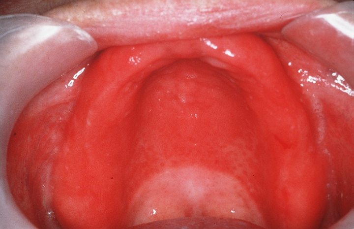
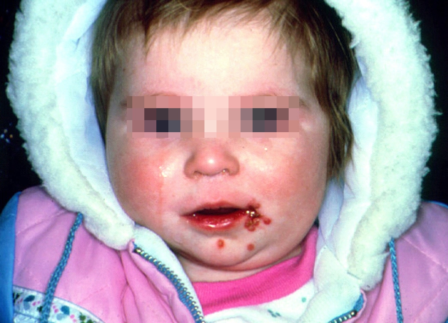
Primary herpetic gingivostomatitis:
Oral Hairy Leukoplakia:

Authors: R. Chigurupati BDS, SS Raghaven PhD, D Studen-Pavlovich DMD
Reference: Pediatric Dentistry 18:2 1996
Purpose: academic review of oral manifestations in pediatric HIV patients
Summary: Pediatric immune systems and developing organ systems are more susceptible to HIV infection and thus degrade more rapidly than adults. Oral lesions are often among the first manifestations of HIV infection due to the number of microorganisms present in the mouth which thrive during immunosuppression. Children can become infected in utero (which brings on the most rapid onset of symptoms, during parturition, breastfeeding, from blood transfusions and other risks. AIDS incidence among African Americans and Hispanic children are 17 and 7 times higher respectively. In particular, the immunosuppression caused by HIV infection can lead to opportunistic fungal and bacterial infections, a propensity toward malignancy, lymphoid interstitial pneumonitis and thrombocytopenia. Encephalitic caused by HIV can result in cognitive, behavioral and motor deficits. Half of all infected infants become symptomatic in the first year of life. Symptoms likely to be seen by dentists: pulmonary lymphoid hyperplasia, salivary gland enlargement, pyogenic bacterial infections such as otitis media, developmental delay and dysmorphic craniofacial features. Children with T cell deficiencies tend to have more oral mucosal candidiasis, HSV and recurrent apthous ulcerations. Children with another type of immunosuppression such as impaired phagocytosis will more often have progressive periodontal disease and parotitis.
ORAL LESIONS: Candidiasis has been documented as the most frequently occuring oral manifestation in HIV infected children with a prevalence ranging from 20-72%. It presents as a creamy white pseudomembranous plaque, erythematous patches, angular chelitis or as non-scrapable hyperplastic plaques. Erythematous or atrophic form appears as flat or raised red patches noted most often on the dorsum of the tongue, palate and buccal mucosa. In infants and small children candidal lesions can be treated by swabbing with nystatin/gentian violet or administeringnystatin pastilles or clotrimazole troches. Antifungals should continue for 1-2 weeks after resolution fo symptoms. Severe cases may be treated by suppressive maintenance antifungal therapy and may be managed by ketaconazole.
You may also encounter parotid enlargement (firm and non-tender, often accompanied by xerstomia), herpetic stomatitis (common, more severe and persistent than in healthy children and will usually recur 2-3 times in less than a year), oral hairy leukoplakia (rare ~2%), petechiae, apthous stomatitis, linear gingival erythema, cervical lymphadenopathy.
It is important to keep track of your HIV patients viral loads, CD4 and platelet counts to anticipate and guide parents and patients as to risk.
Review: A good review of common pediatric soft tissue lesions which may suggest HIV involvement.
Erythematous candidiasis:
Pseudomembranous candidiasis:
Primary herpetic gingivostomatitis:
Oral Hairy Leukoplakia:
Hepatitis B Virus Infection 2/26/10
Department of Pediatric Dentistry
Resident’s Name:Murphy Program: Lutheran Medical Center - Providence
Article title: Hepatitis B Virus Infection
Author(s): Cottone DMD, James. Raghunath Puttaiah, BDS, MPH
Journal: Dental Clinics of North America
Year. Volume (number). Page #’s: 1996. 40(2). 293-307
Major topic: HBV
Minor topic(s): Implications in dentistry
Main Purpose: Review HBV and its occurrence/incidence in the dental setting
Overview of method of research: Review
Findings: Hepatitis, or inflammation of the liver, is can be caused by many things, including viruses, certain diseases, and drug reactions. HBV is one of these viruses. When considering infection control protocols in the dental setting, HBV is the target disease. First described in 1965, HBV is a major cause of acute and chronic liver infection, cirrhosis, and primary hepatocellular carcinoma worldwide. The frequency of HBV varies workdwide, with 90% of the 300 million carriers being in underdeveloped countries (there are over 1,000,000 carriers in the US alone). Clinical signs and symptoms of acute HBV infection include some combination of anorexia, malaise, nausea, vomiting, abdominal pain, jaundice, skin rashes, athralgias, and arthritis. The incubation period of HBV is long…45-160 days. Sequelae to HBV infection could be asymptomatic, symptomatic carrier state, cirrhosis, acute hepatitis infection, primary liver cancer, or death.
HBV can be transmitted both percutaneously and non-percutaneously. Percutaneous transmission is from some kind of stick with a sharp instrument/needle. Non perc. Includes transfer of bodily secretions such as saliva, blood, and cervicular fluid. Transmission of HBV during dental dental treatment occurs primarily in the horizontal mode among staff and patients, mostly from patient to provider and less likely from provider to patient. The risk of HBV is more a factor of exposure to blood than to general patient contact. Intraorally the greatest concentration of HBV is at the gingival sulcus.
Thanks to awareness and immunization, the HBV carrier rate for dentists is only .4%. Dentists who are nonimmune are three times more likely to aquire HBV, and nonimmune specialists are at six times the risk. Oral surgeons have the highest carrier rate at 38.5%. Sorry Dr. Sam!! When compared with HIV, we are 57 times more likely to become infected with HBV.
Vaccines for HBV include a plasma derived vaccine and a recombinant DNA vaccine.
Key points/Summary: As always, proper infection control and universal precautions should always be used for every patient. It’s important that you educate your staff about HBV and other job hazards associated with dentistry. Making sure you and your staff are properly vaccinated, and are up to date with your vaccinations is paramount.
Assessment of Article: Good article….nice review of HBV and it’s implications for us. No shenanigans yo.
Oral soft tissue manifestations and CD4 lymphocyte counts in HIV-infected children
Department of Pediatric Dentistry
Lutheran Medical Center
Date: 02/26/2010
Article title: Oral Soft Tissue Manifestations and CD4 Lymphocyte Coutns in HIV-infected Children
Author(s):Howell, Jandinski, Palumbo, SHey, Houpt
Journal: Pediatric Dentistry
Volume (number): 18:2
Month, Year: 1996
Major topic: HIV related oral manifestations in children.
Minor topics: Realtionship of CD4 lymphocyte levels to the presence of oral soft tissue lesions.
Type of Article: Clinical Investigation
Main Purpose: Investigate the prevalence of oral soft tissue lesions associated with HIV
Overview of method of research: 60 HIV infected children received comprehensive oral examinations. Candida, pseudomembranous and erythematous were grouped with angular cheilitis; Herpes was diagnosed by clinical response to Acyclovir. Modified gingival probings were done. Perio disease diagnosis was based on it’s level of aggression--yikes.
CD4 counts were obtained within 3 months of the oral exams and adjusted for patient age.
Findings:
Even well managed children with HIV often have significant oral disease, Gingivitis in HIV children is associated with (guess what?!) poor oral hygiene, and soft tissue lesions were associated with low CD4 counts.
Key points/Summary :
Because so many HIV pts have oral disease, its presence could be used a a diagnosis factor. Linear gingival erythema was found in 38% of the kids and it is a known manifestation of HIV in adults--that and mustaches.
Children experience less HIV associated periodontitis.
Assessment of article:
This kind of seems like common sense at this point, but it was a good article. Also this study was done 20 years ago (published 5 years later) and our knowledge of HIV has really increased since then.
Monday, February 22, 2010
Oral Manifestations of Pediatric Human Immunodeficiency Virus Infection: A Review of the Literature
Dan Boboia 2/26/10 Lit. Review
Title: Oral Manifestations of Pediatric Human Immunodeficiency Virus Infection: A Review of the Literature
Author: Kline et al
Type of Article: Review
Purpose: To describe the varied oral manifestations of pediatric HIV infections
Erythematous Candidiasis: inconspicous erythematous changes of the palate and dorsum of the tonugue; buccal mucosa involved occasionally; vesicles seen in severe cases; more common among children with low CD4 lymphocyte counts or symptomatic HIV disease than among those with normal counts or no symptoms.
Hairy Luekoplakia: white or gray lesions along the lateral margins of the tongue; lesions appear hairy when affected mucosa is dried; not specific for HIV infection but common for profound immunosuppresion; related to presence of EBV within the oral mucosal epithelium.
Oral Kaposi’s Sarcoma: blue, purple, or red flat or raised patches / nodules, often involving the palate.
Periodontal Disease: several forms of perio are associated with HIV infection; linear gingival erythema has fiery red band along the margin of the gingival; necritizing ulcerative gingivitis show destruction of one or more of the interdental papillae.
Oral Ulcers: lesions often are severely debilitating because they interfere with chewing, speaking, and swallowing.
Cytomeglovirus: another herpes group virus, cam produce large palatal or pharyngeal ulcers resembling major aphthous ulcers as well as changes resemble HIV-associated periodontal disease
Non-Hodgkins lymphoma: is the other form of malignancy diagnosed commonly in adults with AIDS
Sunday, February 21, 2010
Hepatitis C Virus Infection; A review and implications for the dentist
Resident: Roberts
Date: 2/26/10
Article title: Hepatitis C Virus Infection; A review and implications for the dentist
Author: Steven D. Vincent
Journal: Eastman Dental Institute for Oral Health Care Sciences, University of London
Volume: 86 Pages 8-22
Year: 1997
Discussion: Hepatitis C Virus is an RNA virus that is present throughout the world. The virus has many variations and frequently mutates due to poor replication, thus a vaccine or postoperative prophylaxis is far from reaching the marketplace. The virus is mainly transmitted through blood contact, though a large percentage of carriers have an unknown origin. The virus causes chronic hepatitis resulting in cirrhosis and hepatocellular carcinoma in a large majority of its host( after 20 – 30 years of being infected). Interferon alpha is currently the drug of choice but is only effective in about 25% of patients. The most common oral manifestation of the drug is lichen planus or sialadenitis. There have been few reports of noscomial transmission of HCV; however, the prevalence of HCV infection among dental HCW’s is similar to that in the general population.
Assessment: I was left high and dry after the article suggested that is was geared towards dentist. There wasn't a whole lot of information you could really hang your hat on for clinical relevance.
Friday, February 19, 2010
asthma and erosion
Resident’s Name: Joanne Lewis Date: February 19, 2010
Article title: Is there a relationship between asthma and dental erosion? A case control study
Author(s): Al-Dlaigan, Y. H., et al
Journal: International Journal of Pediatric Dentistry
Volume (number): 12
Date: 2002
Major topic: asthma and tooth erosion
Type of Article: research article
Main Purpose: to determine if there is a relationship between asthma and tooth erosion
Overview of method of research: The study consisted of 60 children divided into 3 groups as follows: group 1 – control group of 20 children with no history of asthma or other health problems. Group 2 – asthma group – 20 children with a history of asthma requiring long term medication. Group 3 – erosion group – 20 children referred due to significant erosion, no history of asthma. All children were examined clinically and classified as having low, moderate, or severe erosion. Information about medical history and dietary intake was collected for each child through a standard questionnaire. Unstimulated and stimulated saliva samples were collected from the children – the salivary pH and buffering capacity was measured. The data was analyzed using SPSS.
Findings: None of the children in the control group had severe erosion. In the asthma group, 65% had moderate and 35% had severe erosion. In the erosion group, 65% were classified as having severe erosion, with the majority of the erosion occurring on palatal surfaces, unlike in the control or asthma group. In the asthma and erosion groups there were more children who reported a history of indigestion, vomiting, heartburn and stomach problems. Dietary questions showed a high intake of soft drinks in all three groups of children; however, children in the erosion group did have a diet with more acidic components. There was no statistically significant difference noted in salivary flow rates, salivary pH, or buffering capacity between the 3 groups.
Key points/Summary: Conditions such as vomiting, heartburn, and stomach problems were more common in the erosion and asthma groups. Other studies suggest a definite association between gastroesophageal reflux and asthma. The most consistent dietary risk factor related to erosion is the consumption of soft drinks, carbonated beverages, and sports drinks.
Assessment of article: I thought this study was structured oddly and tried to look at too many variables. Results were not surprising.
Thursday, February 18, 2010
02/19/2010 Treatment of children for primary acute herpetic gingivostomatitis with lactobacillus in aqueous suspension
Resident: J. Hencler Date: 02/19/2010
Article title: Treatment of children for primary acute herpetic gingivostomatitis with lactobacillus in aqueous suspension
Author(s): William F. McCord, DDS
Journal: Pediatric Dentistry: December, 1988 – V 10, #4, Pg 307-308
Major topic: Treatment of primary acute herpetic gingivostomatitis (PAHG)
Type of Article: Clinical trial
Main Purpose: To evaluate the effectiveness of a lactobacillus aqueous preparation in the treatment of PAHG.
Overview of method of research: Study was performed in a private dental office and included 15 males and 18 females ranging from 2-10 years old. Treatment prescribed for each child included a stabilized aqueous suspension of lactobacilli. Parents were instructed to have children swish 1 tsp of the medication in their mouth for as long as possible b/f swallowing and repeat 4x for 7 days.
Findings: All patients experience total relief of pain w/in 72 hrs and all symptoms including lesions and inflammation disappeared w/in 7 days.
Key points in the article discussion: Peak incidence of PAHG is around 2 yo. CC is extreme pain and soreness of the mouth so severe that the child has difficulty eating, drinking, and swallowing. One major problem associated w/ PAHG is dehydration. Results showed relief of pain and lesion healing may have occurred in a shorter period of time than with use of alternate forms of lactobacillus or when lesions remain untreated. The mechanism in which lactobacillus suspension produces a therapeutic response in patients is unknown. It is important to note that this therapeutic approach employs viable organisms of one type (lactobacillus) to combat viable organisms of another type (herpes simplex virus) by acting as a virostatic agent. There is no evidence from this study that the lactobacilli were virostatic other than clinical observation demonstrating cessation of pain, disappearance of lesions, the ability of the child to swallow more easily, the improvement of malaise, and reduction in fever.
Summary of conclusions: This clinical trial presents preliminary evidence that lactobacilli in a liquid form greatly aids in the relief of pain and other symptoms of PAHG. In this study, aqueous lactobacillus preparation showed a high degree of palatability, ease of administration, and may be useful in treating herpetic lesions.
Assessment of article: Small sample size with good results. This was a very interesting article and very easy to read. At the time it was published more research was needed on this tx regimen.
Effect of dental treatment on lung function
Brian Schmid DMD 02/19/10
Authors: Tanya Matthew BDS, Paul S Casamassamo DDS, Stephen Wilson MA PhD.
Reference: JADA Vol 129, August 1998
Main topic: lung function in asthma patients undergoing dental treatment.
Summary: 5-19% of children have some some form of bronchial asthma. The most common triggers for asthmatic attacks include: cigarette smoke, physical exercise, viral and bacterial infections, weather changes including cold temperatures, air pollutants and other allergens and also emotional stress. It has been recommended that dentists limit theur manipulatin of the hyperactive airway during fluoride treatment, radiography and use of cotton rolls. Also, prophylactic use of asthma medication. A semi-reclined or upright can also be used. It is estimated that 40% of attacks are caused by emotional stress. There are also several documented cases of allergies to dental materials, so a rubber dam is recommended to avoid exposure and particularly misting of tooth structure and restoration materials which may irritate the sensitive airway.
57 patients between 6-18 y.o. were chosen and had their lung function tested before, during, immediately post operatively and 30 minutes post operatively for prophylaxis and/or operative visits. Questionnaires were also given to their parents to determine severity of disease, control for several factors and to elucidate how their childs asthma affects their everyday life. All were scheduled after 1pm to control for Circadian changes. The mean patient was 11.8 y.o. and split almost equally for male and female. Fourteen patients (18.8%) showed a clinically significant decrease immediately post-operatively and 12 (16.2%) showed decreased lung function 30 minutes post operatively. Immediate post op and 30 minute post op lung function decrease were associated and predictive. Baseline lung function was the best predictor of post operative lung function. Since spirometry is unliklely to be performed in the dental setting, a thorough history is the best predictor of how sensitive the patient is to attacks and to distinguish common triggers. Severity of asthma and time since last hospital visit were not correlated to incidence of decreased lung function. Clinically 1 in 10 patients had decreased lung function and in this study no clinical variables pertaining to asthma were able to predict a decrease in lung function due to routine dental treatment.
Evaluation: This is a well organized study and reinforces the importance of taking good histories and applying their suggestions clinically.
Role of the pediatric dentist in optimization of childhood immunization
Resident: Roberts
Date: 2/19/10
Article title: Role of the pediatric dentist in optimization of childhood immunization
Journal: American Academy of Pediatric Dentistry
Year: 1996
Discussion:
The American Academy of Pediatric Dentistry stated that “pediatric dentists should consider immunization status as part of the periodic medical history for their patients and should encourage parents to seek appropriate immunization for their children.”
Stats:
- Only ¾ of children have received all the recommended vaccines by 35 months of age.
- Lowest immunizations are associated with poverty(58.7%), African- American(61.8%), non-white races(58.4%), and urban residence(62.1%).
- Highest immunization records are found in those living above the poverty line(70.5%), white race(68.4%), and suburban(71.4%), and rural residents(66%).
- States with highest % of vaccinations include: Vermont (88%), Connecticut (86%), and Hawaii (86%), and low levels are reported in Michigan (61%), Idaho(64%), Missouri(64%).
The need for timely immunization is emphasized by the fact that 17,000 - 23,000 cases of preventable infectious diseases are reported annually to the CDC. The largest percentage of these cases are due to hep. B(63%), pertussis (25%), mumps(8%), and Haemophilus influenza(6%). Children younger than 5 years old represent 20 % of these cases.
Many parents remain uninformed that 16 immunizations are currently required during the first 18 months, with an additional four booster vaccines necessary from age 4 – 16 years. The CDC estimates that more than 2 million kids between the ages of 19 to 36 months are likely inadequetly vaccinated. Dentist are in a an opportune spot to inform parents and encourage vaccinations because they are exposed to children from the earliest of ages.
When a dentist first come across a child that has not been vaccinated properly, they should be referred to a local pediatrician or PCP. If they do not have one or lack the financial means to see one, then they can be referred to a local public clinic where immunizations are done at low or no cost to the patient. The American Academy of Pediatrics has established a National Immunizaion Information Hotline (800-232-2522) on where these clinics can be found.

Department of Pediatric Dentistry
Resident’s Name:Murphy Program:Lutheran Medical Center - Providence
Article title: Resurgence of TB in children
Author(s): Stake MD, Jeffrey.
Journal: The Journal of Pediatrics
Year. Volume (number). Page #’s: 1992. 120. 839-851
Major topic: TB in children
Minor topic(s): TB and the dental setting
Overview of method of research: Clinical review
Findings: The clinical expression of infection due to Mycobacterium tuberculosis differs substantially in children compared with adults. There are three basic stages of the disease: exposure, infection, and disease. Children infected prior to age 4 have a very high rate of developing immediate clinical or radiographic manifestations or both, but are unlikely to develop reactivation disease in adulthood. In contrast, children infected in preadolescence or adolescence are more prone to developing more severe adult-type pulmonary tuberculosis soon after infection or in adulthood. The most common symptoms associated with TB are cough, fever, wheezing, and failure to gain weight. Clinical signs of TB are surprisingly meager, but rales and wheezes over the affected lung field are most common.
Diagnosis
The Mantoux TST(aka PPD), which uses five tuberculin units of purified protein derivative, is the standard method for detecting infection by M. tuberculosis. The reaction is measured as millimeters of induration after 48 to 72 hours. Since TST is the only way to determine asymptomatic infection by M. tuberculosis, the false-negative rate cannot be calculated. A negative TST does not rule out TB disease in a child. If the test is positive, a chest x-ray is taken to either confirm on deny infection. TB in chest radiographs show enlargement of hilar, mediastinal, or subcarinal lymph nodes and lung parenchymal changes. The role of bronchoscopy in evaluating children for TB is controversial.
Treatment
A 6-mo regiment consisting of isoniazid(INH), rifampin, and pyrazinamide given for 2 mo followed by isoniazid and rifampin for 4 mo is the preferred treatment for patients with fully susceptible organisms who adhere to treatment. Ethambutol (or streptomycin in children too young to be monitored for visual acuity) should be included in the initial regimen until the results of drug susceptibility studies are available, unless there is little possibility of drug resistance
Dental Treatment and TB
Because a person with latent TB is not infectious, he or she can be treated in the dental office under standard infection control precautions.2
Any patient with symptoms suggestive of active TB disease should be removed from the area of other patients or staff, instructed to wear a surgical or procedure mask, assessed for the urgency of their dental care and promptly referred for medical care. Standard precautions are insufficient to prevent transmission of the bacterium. Elective dental treatment should be deferred until the patient has been declared non-infectious by a physician. Urgent dental care for a person with suspected or active TB should be provided in a facility that has the capacity for airborne infection isolation and has a respiratory protection program in place. OSHA describes a standard for respiratory protection, which should be consulted if setting up a program. When treating a patient with active TB, dental health care personnel should use respiratory protection (e.g., fitted, disposable N-95 respirators). Standard surgical face masks are not designed to protect against TB transmission. The CDC recommends that dental office personnel receive training and education on M. tuberculosis and TB disease that emphasizes the increased risks posed by an undiagnosed person with TB disease in a dental-care setting and the specific measures to reduce this risk. The CDC also recommends that dental offices perform an annual risk assessment. People in the dental office who have the potential for exposure to M. tuberculosis through air space shared with persons with infectious TB disease (which essentially means all personnel) receive a two-step baseline tuberculin skin test (TST) at the beginning of employment in low-risk settings, every 12 months in medium-risk settings and every 8–10 weeks in the event of potential ongoing transmission until no further evidence of ongoing transmission is apparent.
Assessment of Article: Good article. Kind of old. I also used www.ada.org for additional info, which was very helpful.
http://images.google.com/imgres?imgurl=http://www.nurse.net/clinical/tb/tb.measure.jpg&imgrefurl=http://www.nurse.net/clinical/tb/&usg=__7LFAfkj00ey1aaUaa9oKE-mSo6Q=&h=320&w=500&sz=21&hl=en&start=8&sig2=41RyTZSjlvuQmjM9p0t6tQ&um=1&itbs=1&tbnid=yzXF_f2uTJC-9M:&tbnh=83&tbnw=130&prev=/images%3Fq%3Dpositive%2Bppd%2Bskin%2Btest%26hl%3Den%26rlz%3D1T4DMUS_enUS317US253%26um%3D1&ei=b7p9S6MuibqVB8-d3MUB
Is Asthma a Risk Factor for Dental Caries? Findings from a Cohort Study
Is Asthma a Risk Factor for Dental Caries? Findings from a Cohort Study
Authors: A.M. Meldrum, W.M. Thomson, B.K. Drummond, M.R. Sears
Journal: Caries Research, March 2001
Reviewer: Kris Hendricks
Major topic: To determine if asthma and caries rate were realted
Findings:
Despite suggestions from the literature that asthmatics may be at higher risk for caries, this study found no relationship between asthma and caries rate.
Method:
Cohort study of 781 subjects, 206 had no history of asthma and the others were divided into groups depending on severity of asthma and medication dependence.
Related Findings:
It has been suggested that asthmatics may be at higher risk for caries based on the following
- asthmatics have altered salivary composition and flow rates due to the presence of specific auto-antibodies to beta-adrenergic recepotors. This is caused by administration of beta agaonist anti-asthmatic drugs
- many asthmatic drugs use lactose or other sugars as a delivery agent
- xerostomia plus lactose can cause decay (typically lactose is less cariogenic than other sugars)
- most inhaled asthma powders have a pH below 5.5
- some asthamtics use cariogenic beverages to alleviate xerostomia
My take:
Decent article. They really didn't look at that many asthamtics using a lot of medication--like 50--so I would guess that a lot of the feared effects of asthmatic drugs would affect a highly medicated population more than the asthmatic who has an inhaler but doesn't use it much.
Friday, February 12, 2010
Cleidocranial dysplasia - treatment of dentofacial abnormalities
Resident’s Name: Joanne Lewis Date: February 12, 2010
Article title: Cleidocranial dysplasia: comprehensive treatment of the dentofacial abnormalities
Author(s): Trimble, Douglas DMD MD, et al
Journal: JADA
Volume (number): 105
Date: October 1982
Major topic: Cleidocranial dysplasia
Type of Article: review article
Main Purpose: to describe a 5 phase surgical and orthodontic approach to correcting the dentoalveolar abnormalities seen in patients with cleidocranial dysplasia.
Key points/Summary: Abnormalities of the dentoalveolar complex in patients with CD include – multiple supernumerary teeth, root form abnormalities, failure of primary teeth to exfoliate, failure of permanent teeth to erupt, poorly developed alveolar processes, hypoplastic maxilla, frontal bossing. Possible treatment options include:
Extraction of all primary and permanent teeth with removable prosthesis – problem: retention of denture is compromised due to the amount of alveolar bone removed during extractions and preexisting alveolar insufficiency.
Extraction of all erupted teeth with placement of removable prosthesis – problem: continued tooth eruption results in ulceration, cysts formation, or osteomyelitis. Retention of the erupted permanent teeth with fabricated of an overlay denture has the same problems.
Restoration with a fixed prothesis – problem: poor abutment availability.
The authors advocate a 5 phase surgical and orthodontic treatment plan aimed at restoring function and esthetics.
Phase 1: extraction of primary and supernumerary teeth with exposure and orthodontic banding of the permanent dentition. Considerations: this phase is ideally begun with the crown and approximately 50% of the roots of the canines and premolars have calcified. Extraction decisions made presurgically may have to be altered during surgery due to ankylosis or fusion of the permanent teeth to supernumerary teeth. Made need to retain the primary second molars until the permanent first molars erupt to preserve an occlusal stop and prevent mandibular overclosure. Flap replacement may be complicated by the bulk of orthodontic appliances. Meticulous OH, irrigation, and antibiotic coverage are needed postsurgically. Placement of ortho bands may require extensive removal of alveolar bone.
Phase 2: orthodontic coordination of the maxillary and mandibular dentitions. Considerations: Arch wire is placed at the time of surgery. Teeth are initially highly mobile; may be 6-9 months after surgery before teeth become stable. Once teeth emerge, subsequent orthodontic treatment is conventional.
Phase 3: surgical correction of the facial-skeletal deformities – Le Forte I, etc. Considerations: conventional osteotomy sites may be altered due to abnormal root formation. Final evaluation regarding facial esthetics cannot be made for at least 12 months.
Phase 4: finishing orthodontics.
Phase 5: supplemental treatment procedures – perio, restorative.
Assessment of article: Concise article that gives a good review of the problems associated with CD as well as some relevant clinical information. A good read.
Thursday, February 4, 2010
Regional odontodysplasia, diagnosis, and description
Department of Pediatric Dentistry
Lutheran Medical Center
Name: Craig Elice Date: January 22, 2010
Article title: Regional Odontodysplasia: a bibliography.
Author(s): Crawford PJM, Aldred MJ.
Journal: J. Oral Pathology Medicine
Volume (number): 18:251-63
Month, Year: 1989
Major topic: Regional odontodysplasia, diagnosis, and description
Type of Article: Review of Literature
Main Purpose: The article provided an overview of the characteristics of regional odontodysplasia, a differential diagnosis, and an expansive review of the literature.
Findings: 109 cases have been described in the literature that primarily affects the coronal part of the teeth, although the enamel, dentin, pulp and follicle have been involved. It occurs more frequently in females. No consistent family history, pregnancy or birth history , or medical history has been noted. The disorder presents generally at the time of eruption of the primary or permanent teeth and the presenting sign is failure, delayed or partial arrested eruption of teeth or abscesses with no known caries. The teeth are often discolored with roughened surface texture. The maxillary arch is more commonly affected and it is usually in a continuous series although skipping has been reported especially the bicuspid teeth. It has also been reported that permanent teeth can be affected and not the primary teeth, although the authors suspect that the primary teeth were affected and were not recognized as such. Radiographically, these teeth appear as “ghost like” with no contrast between enamel and dentin. The pulp spaces are large and frequently have pulp stones, while the roots are shortened with open apices. Histopathology of the enamel, dentin and cementum is variable. Enamel hypoplasia is common with invaginations communicating with the pulp have been reported. Interglobular dentin in the crown of the teeth is a common finding with amorphous areas resembling cementum or calculus. These characteristics were suggested as pathognomonic of odontodysplasia. The roots of affected teeth tend to have a normal structure. Associated pathology with other disorders has been suggested but is inconclusive. The etiology of the condition is also variable. Vascular and neural crest cell theories, viral, traumatic incidents, medications, syndromal involvement, and metabolic disturbances were all mentioned with uncertain relationships. The authors suggest an idiopathic etiology. Treatment is usually conservative and the teeth are best retained whenever possible to preserve the alveolar ridge for future prosthesis. Extactions are indicated in the case of an abscess with no other complications
Key points/Summary: It is important to recognize regional odontodysplasia as a differential diagnosis in order to rule out other more ominous disorders.
Assessment of article: Good but long article which provides comprehensive information about the full discussion of regional odontodysplasia.
Clinical diagnosis and management strategies of amelogenesis imperfecta variants
Resident: Adam J. Bottrill
Date: 05FEB10
Region: Providence
Article title: Clinical diagnosis and management strategies of amelogenesis imperfecta variants
Author(s): Seow, W. Kim
Journal: Pediatric Dentistry
Page #s: pp. 384-393
Year: 1993
Major topic: Amelogenesis Imperfecta
Minor topic(s): None
Type of Article: Topic Summary
Main Purpose: The aim of this study was to analyze the clinical presentations, diagnostic factors and clinical complications of different variants of AI.
Overview of method of research: Patient study and comparison. 32 patients from 17 families.
Key points in the article discussion:
I. AI is a group of inherited disorders primarily affecting dental enamel.
A. Prevalence: from 1:718 to 1:14000 depending on population.
B. Etiology: Alteration of the genes involved in the complex process of enamel formation and maturation.
C. Dx: 1-generalized enamel hypoplasia in primary and permanent teeth. 2-FHx or new mutation 3-absence of systemic factors.
II. Hypoplastic: characterized by deficiency in quantity of enamel (thin enamel or pits and grooves)
A. Pitted (autosomal dominant): small, pinpoint size pits in rows, localized enamel loss, normal radiographs
1. Clinical Problems: Mostly esthetic, mild gingivitis
B. Local (autosomal recesive): horzontal bands of pitting or missing enamel, opaque enamel, normal radiographs.
1. Clinical Problems: Esthetic and sensitivity.
C. Smooth, thin (autosomal dominant): thin, hard, smooth, glossy enamel, colored from white to brown, narrow teeth, no contacts, thin enamel on radiograph.
1. Clinical Problems: Esthetics, sensitivity, loss of VDO, better gingival health than other types.
D. Smooth (X-linked): lyonization, vertical ridges and grooves, thin enamel on radiograph. Father can’t trasmit.
1. Clinical Problems: Esthetics, loss of VDO, gingivitis.
E. Rough (autosomal dominant): thin, hard, rough enamel, minimal contacts, thin enamel on radiograph
1. Clinical Problems: Esthetics, sensitivity, loss of VDO, gingivitis.
III. Hypocalcified: characterized by enamel that is insufficiently mineralized (soft, discolored enamel)
A. (autosomal recessive/dominant): soft, opaque, yellow enamel, chips away easily, adequate contacts, MINIMAL contrast between dentin and enamel on radiograph.
IV. Hypomaturation: characterized by abnormalities in the maturation stages of dev (opaque, chalky enamel)
A. (autosomal recessive/x-linked recessive): opaque enamel, normal contacts, mild open bite, lack of contrast between enamel and dentin on radiograph, similar appearance to fluorosis,
Assessment of article: Basic and informative. Shenanigans were not found here.
Tricho-Dento-Osseous Syndrome
Resident’s Name: Joanne Lewis Date: February 5, 2010
Article title: Analysis of the Tricho-Dento-Osseous Syndrome Genotype and Phenotype
Author(s): Wright, J. Tim DDS MS, et al
Journal: American Journal of Medical Genetics
Volume (number): 72
Date: 1997
Major topic: Tricho-Dento-Osseous Syndrome
Type of Article: scientific article
Main Purpose: to characterize the phenotype of TDO in 3 previously undescribed kindreds in North Carolina and to test genetic linkage to specific candidate regions.
Overview of method of research: 53 individuals (33 affected individuals and 20 non-affected individuals) were recruited to participate in the study. Detailed family histories as well as medical and dental histories were obtained for each individual. Participants were evaluated clinically and radiographically. Blood was drawn for genetic linkage analysis.
Findings: Curly/kinky hair at birth was found in 85% of affected individuals, with 46% retaining the curly hair phenotype after infancy. The TDO teeth were small (due to enamel hypoplasia) and often had a slight yellow-brown coloration. Pitting and/or furrowing of the enamel was evident in 63% of the teeth. Dental abscesses were reported by 77% of individuals with TDO, as opposed to 25% of unaffected persons. Taurodontism was present in some degree in all affected individuals. Skeletal manifestations in TDO varied substantially and included an increase in cranial bone density, calvarial diploe obliteration, and a lack of visible mastoid pneumatization.
Key points/Summary: Curly hair at birth, enamel hypoplasia, and taurodontism are highly penetrant yet clinically variable components of TDO. The ABO, Kell, and Gc loci previously suggested to be linked to TDO were excluded as candidates in this TDO population.
Assessment of article: Provides support for the “soft-tooth” theory.
Amelogenesis Imperfecta (AI), dentinogenesis inperfecta (DI), and dentin dysplasia (DD) revisited: problems in classification
Resident: J. Hencler
Date: 02/05/2010
Article title: Amelogenesis Imperfecta (AI), dentinogenesis inperfecta (DI), and dentin dysplasia (DD) revisited: problems in classification
Author: C. J. Witkop Jr.
Journal: J Oral Pathol 1989: 17: 547-553
Major topic: AI, DI, DD
Type of Article: Review
Main Purpose:
Update classification and prevalence of AI and discuss problems with the classification of inherited dentin defects.
Discussion:
AI should is limited to those inherited, congenital defects that only affect enamel formation and not accompanied by morphogenic or metabolic defects in other body systems other than tooth form or eruption. AI classification based on clinical exam is problematic b/c the same clinical features may have several modes of inheritance. The expression of AI can vary greatly from very mild forms to severe. AI prevalence differed in many studies at the time of this publication so was still debated. Now the estimated incidence of AI includes 1 in 14000, 1 in 8000, and 1 in 4000. 14 subgrps of AI with multiple inheritance patterns are known which complicates diagnosis. The 4 categories of AI are described according to the stage of tooth development in which each is thought to occur.
DI is an inheritable dental defect of pre-dentin matrix that results in amorphic, disorganized, and atubular circumpulpal dentin. DI is classified into 3 types. Type 1 occurs with osteogenesis imperfect (OI), Type 2 is also known as hereditary opalescent dentin. Type 3 is a rare type found only in a tri-racial isolated grp in Maryland called the Brandywine population and is proposed to be a different expression of the same type 2 gene.
DD represents a grp of inherited dentin disorders resulting in defects involving circumpulal dentin and root morphology. 2 Types are described. Type 1 has normal primary and perm crown morph but with amber translucency. Roots are short and sharply constricted. Primary teeth have obliterated pulps and both dentitions exhibit multiple radiolucencies and absent pulp chambers. Type 2 involves amber colored primary teeth closely resembling DI types 1 and 2. Perm teeth appear normal, but radiographically show thistle tube shaped pulp chambers with pulp stones. DD type 2 has been argued to be a type of DI on the basis of clinical, radiographic, histologic, and alterated structural protein similarities.
Assessment of article:
Awful, it was hard to understand and written in way too much genetic detail. Unless you enjoy an article that debates numerous intricate genetic differences that determine a complicated classification system, this article may not be for you. The article did do a good job going into each subtype of AI but lacked info on DI and DD.
Retrospective study of children with hypophosphatasia with reference to dental changes 2/5/10
Department of Pediatric Dentistry
Resident’s Name: Murphy Program: Lutheran Medical Center - Providence
Article title: Retrospective study of children with hypophosphatasia with reference to dental changes
Author(s): Lundgren, Ted, Otto Westphal, Per Bolme, Thomas Moderer, et al
Journal:
Year. Volume (number). Page #’s: Jan 1991. 357-364
Major topic: Hypophosphatasia
Minor topic(s): Dental implications of HP
Main Purpose: Discuss the effects HP had on 17 children
Overview of method of research: Eval. Of 17 children
Findings: Hypophosphatasia(HP) is lack of the bone/liver/kidney isoenzyme alkaline phosphatase(AP). There are infantile, childhood, and adult forms of the disease. There is a hereditary component. Clinical expressions range from
1. intrauterine death
2.severe skeletal hypomineralization/numerous fractures
3. Leg symptoms such as hip, knee, ankle problems, trouble walking, valgus(the outward angulation of the distal segment of a bone or joint) feet o r knees, limited motion, limping, and no babinski sign
4. Premature loss of one or more primary incisors with no hx of trauma
5. Insufficient weight gain and GI issues
6. Changes in skull sutures and fontanelle
7. Eye anomalies in the retina and sclera
8. Marginal bone loss in anterior mandible region
9. Enlarged pulp cavities
10.Phenylethylamine (PEA) in the urine
Due to its early oral manifestations, dentists are usually the first ones to suspect and/or diagnose the disorder. A typical scenario is a child presents after losing his 4 mand. incisors with no history of trauma. The most common finding on dental radiographs is marginal bone loss in the mandibular anterior region, along with cementum aplasia.
Key points/Summary: Measuring the PEA in the urine is the best test for HP. An exam of the legs and radiographic exam of the long bones is essential. A Dental exam with special attention to premature loss of primary teeth and assessment of anterior mandibular bone an be an early predictor.
Assessment of Article: Good article. Quick and dirty review of HP and it’s oral manifestations
http://upload.wikimedia.org/wikipedia/commons/0/0a/Valgus.jpg
Wednesday, February 3, 2010
Hereditary Defects of Enamel
Department of Pediatric Dentistry
Lutheran Medical Center
Date: 02/05/2010
Article title: Hereditary Defects of Enamel
Author(s): J Tim Wright
Journal: Text: Dentin Enamel: Formation to Destruction
Volume (number):
Month, Year: 1995
Major topic: Enamel defects
Minor topics: Syndromes associated with enamel defects
Type of Article: Text, educational
Main Purpose: To Present the current knowledge of hereditary enamel disorders
Overview of method of research: Text based on a review of the literature
Findings:
This text has a ton of information. It is an important read, but not important for me to copy again. After board review, my feeling is that by knowing the information in Table 1, you’ll be well prepared for the exam.
I reproduced it here and added some pertinent information. I did not add any histology as it is probably not relevant to what we need for the boards.
Key points/Summary :
- Syndromic conditions
- Metabolic disorders
- Mucopolysaccharidosis type IV (mucopolysaccharides accumulate around developing teeth)
- Hypophosphatasia (abnormal cementum, early primary tooth exfoliation, pitting)
- Vitamin D-dependent rickets
- Vitamin D-resistant rickets
- Pseudohypoparathyroidism
- Ectodermal/mesenchymal disorders
- Ectodermal dysplasias
- Epidermolysis bullosa, junctional type (skin fragility, blistering, dysplastic enamel)
- Tricho-dento-osseous syndrome
- Icontinentia pigmenti
- Tuberous sclerosis
- Oculo-dento-osseous syndrome
- Nonsyndromic Conditions
- Amelogenesis imperfecta (usually only for defects exclusively in enamel, the 3 major groups are listed)
- Hypoplastic type
- Hypocalcified type
- Hypomaturation type
Assessment of article: This was a good read. I think that everyone should be sure to read this over as it helps us better understand teeth generally.
Subscribe to:
Posts (Atom)