Authors: R. Chigurupati BDS, SS Raghaven PhD, D Studen-Pavlovich DMD
Reference: Pediatric Dentistry 18:2 1996
Purpose: academic review of oral manifestations in pediatric HIV patients
Summary: Pediatric immune systems and developing organ systems are more susceptible to HIV infection and thus degrade more rapidly than adults. Oral lesions are often among the first manifestations of HIV infection due to the number of microorganisms present in the mouth which thrive during immunosuppression. Children can become infected in utero (which brings on the most rapid onset of symptoms, during parturition, breastfeeding, from blood transfusions and other risks. AIDS incidence among African Americans and Hispanic children are 17 and 7 times higher respectively. In particular, the immunosuppression caused by HIV infection can lead to opportunistic fungal and bacterial infections, a propensity toward malignancy, lymphoid interstitial pneumonitis and thrombocytopenia. Encephalitic caused by HIV can result in cognitive, behavioral and motor deficits. Half of all infected infants become symptomatic in the first year of life. Symptoms likely to be seen by dentists: pulmonary lymphoid hyperplasia, salivary gland enlargement, pyogenic bacterial infections such as otitis media, developmental delay and dysmorphic craniofacial features. Children with T cell deficiencies tend to have more oral mucosal candidiasis, HSV and recurrent apthous ulcerations. Children with another type of immunosuppression such as impaired phagocytosis will more often have progressive periodontal disease and parotitis.
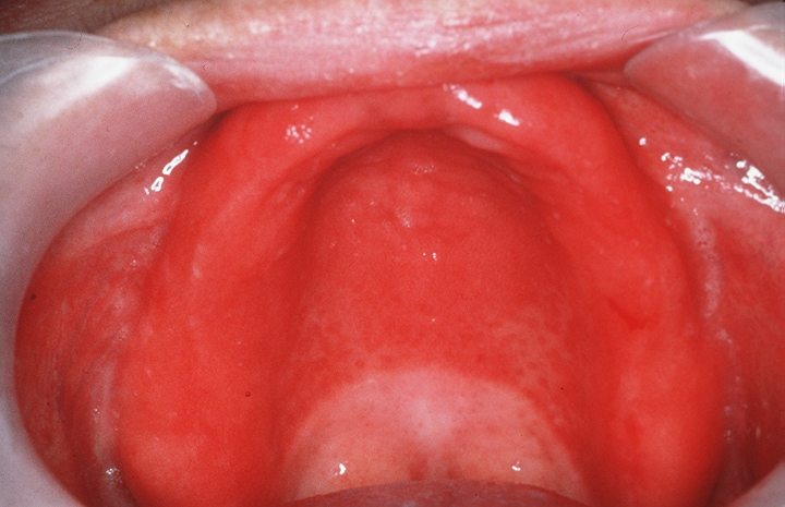
ORAL LESIONS: Candidiasis has been documented as the most frequently occuring oral manifestation in HIV infected children with a prevalence ranging from 20-72%. It presents as a creamy white pseudomembranous plaque, erythematous patches, angular chelitis or as non-scrapable hyperplastic plaques. Erythematous or atrophic form appears as flat or raised red patches noted most often on the dorsum of the tongue, palate and buccal mucosa. In infants and small children candidal lesions can be treated by swabbing with nystatin/gentian violet or administeringnystatin pastilles or clotrimazole troches. Antifungals should continue for 1-2 weeks after resolution fo symptoms. Severe cases may be treated by suppressive maintenance antifungal therapy and may be managed by ketaconazole.
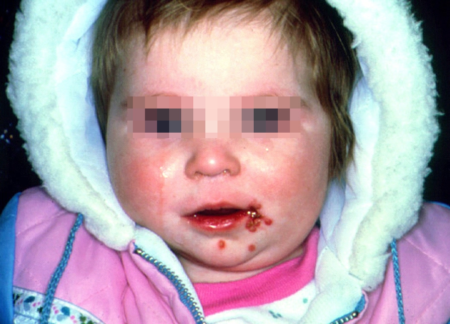
You may also encounter parotid enlargement (firm and non-tender, often accompanied by xerstomia), herpetic stomatitis (common, more severe and persistent than in healthy children and will usually recur 2-3 times in less than a year), oral hairy leukoplakia (rare ~2%), petechiae, apthous stomatitis, linear gingival erythema, cervical lymphadenopathy.
It is important to keep track of your HIV patients viral loads, CD4 and platelet counts to anticipate and guide parents and patients as to risk.
Review: A good review of common pediatric soft tissue lesions which may suggest HIV involvement.
Erythematous candidiasis:
Pseudomembranous candidiasis:

Primary herpetic gingivostomatitis:
Oral Hairy Leukoplakia:

No comments:
Post a Comment