Resident: Adam J. Bottrill
Date: 26FEB10
Region: Providence
Article title: Herpetic gingivostomatitis and teething difficulty in infants
Author(s): King, David et. al.
Journal: Pediatric Dentistry
Page #s: pp. 82-85
Year: March/April 1992
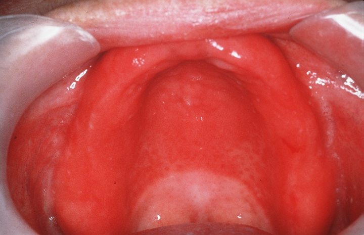
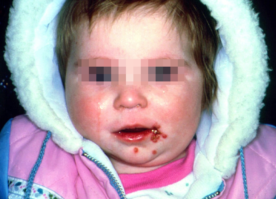
Major topic: Primary herpetic gingivostomatitis (PHG)
Minor topic(s): Teething
Type of Article: Retrospective investigation
Main Purpose: The aim of this study was to determine whether PHG may be responsible for signs and symptoms commonly attributed to teething in infants.
Overview of method of research: Patient study and comparison. 20 infants with parental diagnosis of “teething difficulty.” 20 infants serving as controls.
Key points in the article discussion:
I. Oral swab obtained from the infants and processed to determine presence of HSV.
A. Temp and oral status recorded.
B. 9 subjects positive for HSV
C. 7 of 9 had temperature
D. All 9 had varying degrees of visible oral infection
E. Of the remaining 11, 5 had temperatures and none had visible infeciton (otitis media, vericella).
F. The control group was negative for HSV, temp AND visible infection.
II. Results: HSV should be included in differential Dx of patients with CC of “teething difficulty”
A. Finding of HSV “highly significant” in the DDX of teething difficulty.
B. 80-90% of adults have serum AB’s to HSV. Many don’t remember having it. This could be due to undiagnosed infant PHG.
C. Infants with teething difficulty should be more closely analyzed for PHG.
Assessment of article: Short and Sweet. Negative on the Shenanigans.
Thursday, February 25, 2010
Revised pediatric HIV classification system
Resident: J. Hencler
Date: 02/26/2010
Article title: Revised pediatric HIV classification system
Author(s): AAPD
Journal: Pediatric Dentistry-18:2,1996
Major topic: HIV
Type of Article: Revision
Key points in the article discussion:
In 1994, a new classification system for pediatric HIV replaced the 1987 system (see tables)
Category N (not symptomatic): Includes children who have no signs or symptoms considered to be the result of HIV infection or who have only one of the conditions listed in Category A.
Category A (mildly symptomatic): Children with two or more of the conditions listed below but none of the conditions listed in Categories B and C. (see list)
Category B (moderately symptomatic): Children who have symptomatic conditions other than those listed for Category A or C that are attributed to HIV infection. Examples of conditions in clinical Category B include but are not limited to: (see list)
Category C (severely symptomatic): Serious bacterial infections, multiple or recurrent (i.e., any combination of at least two culture-confirmed infections within a 2-year period), of the following types: septicemia, pneumonia, meningitis, bone or joint infection, or abscess of an internal organ or body cavity (excluding otitis media, superficial skin or mucosal abscesses, and indwelling catheter-related infections). (see list)
Summary of conclusions: New pediatric HIV classification system is simpler. It would be good to have a reference manual if needed.
Assessment of article: This revision of the pediatric HIV classification system is good to be aware of because many signs and symptoms of HIV infection can manifest in the oral cavity. This classification is also pertinent to us as pediatric dentists when consulting with a Pediatrician about a child’s HIV status and severity.
Pediatric AIDs
Department of Pediatric Dentistry
Lutheran Medical Center
Name: Craig Elice Date: February 26, 2010
Article title: Pediatric Aids
Author(s): Chadwick EG, Yogev R.
Journal: Dental Clinics of North America
Volume (number): 42(4) 969-979
Month, Year: August 1995
Major topic: summary of Pediatric Aids
Type of Article: Review of Literature
Main Purpose: The article provided an overview of the HIV and AIDs.
1 million of 13 million infected persons are children with 80% in sub-saharan populaton. The prenatal transmission rate is 20-30%. Approximately 89% of infected children less than 13 years of age were infected by vertical transmission which is increasing while less than 10% are infected through contaminated blood products. African Americans represent 15% of the childhood population yet represent 50 % pf the pediatric AIDs cases. Adolescents between 13 and 19 represent the fastest growing segment of Aids cases. Risk factors vary by gender in adolescents. 45% of males with AIDs are infected by contaminated blood or blood products while females are infected by heterosexual contact. Transmission: In pediatric populations, vertical transmission is most common with 12-20% transmission rates in US and Europe and igher transmission rates in Africa and Haiti. This vertical transmission can occur intrauterine, during delivery, or less often via breast feeding. Transfusion s of infected blood account for 9% of pediatric AIDs and it appears to be diminishing as a result of HIV antibody screening. HIV is rarely isolated from saliva and does not appear to be a real danger of transmission. Pathogenesis: HIV binds to cells with CD$ molecules (T4 lymphocytes) which migrate to lymph nodes where they become activated and proliferate. Patients develop an immune response which suppresses HIV levels yet causes a deterioration of the immune response. This suppression phase is the clinical “Latency phase”. Eventually the immune system deterioration frees the virus to recirculate through many body tissues Clinical Manifestations: The CDC pediatric guidelines include status of disease: mild to moderate to severe signs and symptoms as well as the immune classification based of absolute T-helper cells count or the Percent of CD4 cells. Recurrent bacterial infections, chonic parotid swelling, LIP, and early onsey of progressive neurological deterioration are characteristic of pediatric AIDs. Infections: The most common serious infections affecting greater than50% of infected children are bacteremia, sepsis, and pneumonia. While adults experience reactivation of latent infections like viruses, most pediatric infections are primary including pneumocystis carinii pneumonia (PCP). Oral candidiasis is the most common fungal disease in HIV infected children. Viral infections like JSV, VZV, and CMV are common. Systemic: CNS involvement in vertically infected children is approximately 20-50% and presents as progressive encephalopathy with loss or stagnation of milestones, cognitive deterioration and stunted brain growth. Recurrent respiratory like otitis and sinusitis are common. LIP affects 1/3 of children presenting as lymphoid hyperplasia of the bronchia tubules causing blockage of the alveolar/ capillary interface. PCP is the most common opportunistic infection in pediatric AIDs Cardiac involvement in patients with HIV infection has been thoroughly studied. GI involvement is common with symptoms of chronic and recurrent diarrhea with malabsorption abdominal pain, dysphagia, and failure to thrive (FTT). Chronic liver inflammation is common. Renal disease is an uncommon finding. Skin diseases although present in healthy children, are common in HIV infected children. Recurrent and chronic primary infections with HSV, Zoster, and other viruses are common as well as fungal diseases as well as hyperkeratosis. Malignant diseases are rare in pediatric AIDs. Diagnosis is confirmed using immunoassay for IgG antibody to HIV after 18 months of age, while diagnosis is made by virus culture in infants. Management: Focus is placed on nutritional status of patients and prevention of PCP which is difficult as PCP sometimes precedes the diagnosis of AIDs. Guidelines from the CDC for prophylaxis target infants born to infected mothers or shown to be infected by HIV. Therapy includes use of Zidouridine as the first drug of choice in children who have a reduced CD4 count and symptoms of AIDs. Combinations of other meds like Didanosine and others are being investigated. Prognosis is poor when the patient is symptomatic during the first year of life. Opportunistic infections, encephalopathy also carry a poor prognosis.
Key points/Summary: Describes the pathophysiology of pediatric AIDs.
Assessment of article: Good article but may need an update.
Herpes virus Infections
Department of Pediatric Dentistry
Lutheran Medical Center
Name: Craig Elice Date: February 26, 2010
Article title: Herpes virus Infections
Author(s): Greenberg MS.
Journal: Dental Clinics of North America
Volume (number): 40:2 pages 359-368
Month, Year: April 1996
Major topic: summary of herpes viruses
Type of Article: Review of Literature
Main Purpose: The article provided an overview of the herpes viruses that infect human beings.
Of the 80 known viruses, only 7 Herpes viruses infect humans: HSV I and 2, Varicella Zoster (VSV), Cytomegalovirus (CMV), Epstein- Barr (EBV), HHV 6 and HHV 7. Characteristics that they share include: All viruses cause a primary infection upon first patient exposure and become latent within nuclei of certain body cells for the life of the patient. HSV I, 2, and VZV become dormant in sensory nerve ganglia, CMV in lymphocytes and salivary glands, EB in B-lymphocytes and salivary gland tissues, HHV 6 and 7 in CD4 lymphocytes. They recur as localized symptomatic or asymptomatic recurrent infection., Are transmitted by direct contact via saliva or genital secretions. The EBV can transform cells into maligniancy like nasopharyngeal carcinoma Burkitts lymphomaor B cell lymphoma. HSV 1 and 2: HSV 1 is primarily transmitted by saliva and causes most oral, pharyngeal, eye and CNS infections, while HSV 2 is transmitted by genital secretions and casues genital and anal infections. The incidence of primary infections for HSV 1 is after 6 months of age and peakes between 2-3 years of age. Primary infections are often subclinical but can be characterized by fever, chills, nausea, and lymphadenopathy and followed in 1-2 days by vesicles and ulcers of oral tissues and gingivitis. 20-40% of patients develop recurrent infections which are activated by local trauma or systemic fever or menstrual cycle. Diagnosis is made clinically but viral culture and cytology smears and serology can confirm the diagnosis. CMV is mostly asymptomatic in humans except as a congenital infection called Cytomegalovirus inclusion disease. It is transmitted via birth canal secretions, breast milk, saliva in young children, contaminated blood transfusions, and sexual contact. It is a cause of disease in immunocompromised patients like transplant and AIDs patients. Diagnosis is made by histopathologic studies sowing enlarged owl eye cells. VSV: In 0.3-0.5% of the infected patients, the virus becomes activated presenting as herpes zoster. C-3, T-5, L-1 and 2, are the nerves tracts most commonly infected, but can infect the trigeminal. The symptoms include pain, tenderness and paresthsia along the course of the infected nerve which often precedes vesicles. These vesicles along the nerve tract are a diagnostic sign. In immunocompromised patients, the disease can be life threatening. Acyclovir is effective in shortening the duration and accelerates healing. EBV: Although mild in children, in adolescents and young adults, it can lead to infective mononucleosis. Mono has an incubation period of up to 8 weeks and presents as fever, malaise, pharyngitis, and lymphadenopathy. Diagnosis is based on clinical signs and detection of large lymphocytes in the peripheral blood. Oral lesions include hairy leukoplakia HHV 6: The virus infects CD$ lymphocytes and other white blood cells causing roseola which is self limiting. HHV 7: Usually transmitted by saliva and is found in 80% of adults and 70% if children. NO clinical disorder have been associated with it.
Key points/Summary: Describes 7 human herpes viruses.
Assessment of article: Good article but may need an update.
Oral manifestations of HIV in kids
Resident’s Name: Joanne Lewis Date: February 26, 2010
Article title: Oral findings in asymptomatic (P-1) and symptomatic (P-2) HIV-infected children
Author(s): Agnes Del Toro, DDS, et al
Journal: Pediatric Dentistry
Volume (number): 18(2)
Month, Year: 1996
Major topic: oral manifestations of HIV in children
Type of Article: research
Main Purpose: to assess the relationship between HIV-related oral findings and HIV disease progression in perinatally HIV-infected children.
Overview of method of research: 28 HIV-infected children were selected for inclusion in the study: 13 females, 15 males ranging in age from 2 months to 13.5 years. All of the patients acquired HIV via maternal transmission and all were taking antiretroviral medication at the time of oral examination. 10 of the patients were classified as P-1 (asymptomatic) and 18 of the patients were classifies as P-2 (symptomatic) – CDC classification system.
Findings: The oral manifestations found in this population were pseudomembranous candidiasis, minor aphthous ulceration, delayed dental development, parotid swelling, and petechiae. 11 of the 28 patients (39.3%) had an oral finding; none of the 11 had more than 1 oral finding. 10% of the P-1 patients and 56% of the P-2 patients had an oral finding. The most common oral finding was pseudomembranous candidiasis.
Key points/Summary: Oral findings are common in pediatric HIV infection. Not surprisingly, P-2 patients have a significantly higher occurrence of oral findings than P-1 patients. Oral candidiasis is one of the most common oral complications of pediatric HIV infection.
Assessment of article: Clinically relevant – short and sweet.
Pediatric HIV infection and its oral manifestations: a review
Title: Pediatric HIV infection and its oral manifestations: a review
Authors: R. Chigurupati BDS, SS Raghaven PhD, D Studen-Pavlovich DMD
Reference: Pediatric Dentistry 18:2 1996
Purpose: academic review of oral manifestations in pediatric HIV patients
Summary: Pediatric immune systems and developing organ systems are more susceptible to HIV infection and thus degrade more rapidly than adults. Oral lesions are often among the first manifestations of HIV infection due to the number of microorganisms present in the mouth which thrive during immunosuppression. Children can become infected in utero (which brings on the most rapid onset of symptoms, during parturition, breastfeeding, from blood transfusions and other risks. AIDS incidence among African Americans and Hispanic children are 17 and 7 times higher respectively. In particular, the immunosuppression caused by HIV infection can lead to opportunistic fungal and bacterial infections, a propensity toward malignancy, lymphoid interstitial pneumonitis and thrombocytopenia. Encephalitic caused by HIV can result in cognitive, behavioral and motor deficits. Half of all infected infants become symptomatic in the first year of life. Symptoms likely to be seen by dentists: pulmonary lymphoid hyperplasia, salivary gland enlargement, pyogenic bacterial infections such as otitis media, developmental delay and dysmorphic craniofacial features. Children with T cell deficiencies tend to have more oral mucosal candidiasis, HSV and recurrent apthous ulcerations. Children with another type of immunosuppression such as impaired phagocytosis will more often have progressive periodontal disease and parotitis.
ORAL LESIONS: Candidiasis has been documented as the most frequently occuring oral manifestation in HIV infected children with a prevalence ranging from 20-72%. It presents as a creamy white pseudomembranous plaque, erythematous patches, angular chelitis or as non-scrapable hyperplastic plaques. Erythematous or atrophic form appears as flat or raised red patches noted most often on the dorsum of the tongue, palate and buccal mucosa. In infants and small children candidal lesions can be treated by swabbing with nystatin/gentian violet or administeringnystatin pastilles or clotrimazole troches. Antifungals should continue for 1-2 weeks after resolution fo symptoms. Severe cases may be treated by suppressive maintenance antifungal therapy and may be managed by ketaconazole.
You may also encounter parotid enlargement (firm and non-tender, often accompanied by xerstomia), herpetic stomatitis (common, more severe and persistent than in healthy children and will usually recur 2-3 times in less than a year), oral hairy leukoplakia (rare ~2%), petechiae, apthous stomatitis, linear gingival erythema, cervical lymphadenopathy.
It is important to keep track of your HIV patients viral loads, CD4 and platelet counts to anticipate and guide parents and patients as to risk.
Review: A good review of common pediatric soft tissue lesions which may suggest HIV involvement.
Erythematous candidiasis:

Pseudomembranous candidiasis:

Primary herpetic gingivostomatitis:

Oral Hairy Leukoplakia:

Authors: R. Chigurupati BDS, SS Raghaven PhD, D Studen-Pavlovich DMD
Reference: Pediatric Dentistry 18:2 1996
Purpose: academic review of oral manifestations in pediatric HIV patients
Summary: Pediatric immune systems and developing organ systems are more susceptible to HIV infection and thus degrade more rapidly than adults. Oral lesions are often among the first manifestations of HIV infection due to the number of microorganisms present in the mouth which thrive during immunosuppression. Children can become infected in utero (which brings on the most rapid onset of symptoms, during parturition, breastfeeding, from blood transfusions and other risks. AIDS incidence among African Americans and Hispanic children are 17 and 7 times higher respectively. In particular, the immunosuppression caused by HIV infection can lead to opportunistic fungal and bacterial infections, a propensity toward malignancy, lymphoid interstitial pneumonitis and thrombocytopenia. Encephalitic caused by HIV can result in cognitive, behavioral and motor deficits. Half of all infected infants become symptomatic in the first year of life. Symptoms likely to be seen by dentists: pulmonary lymphoid hyperplasia, salivary gland enlargement, pyogenic bacterial infections such as otitis media, developmental delay and dysmorphic craniofacial features. Children with T cell deficiencies tend to have more oral mucosal candidiasis, HSV and recurrent apthous ulcerations. Children with another type of immunosuppression such as impaired phagocytosis will more often have progressive periodontal disease and parotitis.
ORAL LESIONS: Candidiasis has been documented as the most frequently occuring oral manifestation in HIV infected children with a prevalence ranging from 20-72%. It presents as a creamy white pseudomembranous plaque, erythematous patches, angular chelitis or as non-scrapable hyperplastic plaques. Erythematous or atrophic form appears as flat or raised red patches noted most often on the dorsum of the tongue, palate and buccal mucosa. In infants and small children candidal lesions can be treated by swabbing with nystatin/gentian violet or administeringnystatin pastilles or clotrimazole troches. Antifungals should continue for 1-2 weeks after resolution fo symptoms. Severe cases may be treated by suppressive maintenance antifungal therapy and may be managed by ketaconazole.
You may also encounter parotid enlargement (firm and non-tender, often accompanied by xerstomia), herpetic stomatitis (common, more severe and persistent than in healthy children and will usually recur 2-3 times in less than a year), oral hairy leukoplakia (rare ~2%), petechiae, apthous stomatitis, linear gingival erythema, cervical lymphadenopathy.
It is important to keep track of your HIV patients viral loads, CD4 and platelet counts to anticipate and guide parents and patients as to risk.
Review: A good review of common pediatric soft tissue lesions which may suggest HIV involvement.
Erythematous candidiasis:
Pseudomembranous candidiasis:

Primary herpetic gingivostomatitis:
Oral Hairy Leukoplakia:

Hepatitis B Virus Infection 2/26/10
Department of Pediatric Dentistry
Resident’s Name:Murphy Program: Lutheran Medical Center - Providence
Article title: Hepatitis B Virus Infection
Author(s): Cottone DMD, James. Raghunath Puttaiah, BDS, MPH
Journal: Dental Clinics of North America
Year. Volume (number). Page #’s: 1996. 40(2). 293-307
Major topic: HBV
Minor topic(s): Implications in dentistry
Main Purpose: Review HBV and its occurrence/incidence in the dental setting
Overview of method of research: Review
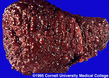
Findings: Hepatitis, or inflammation of the liver, is can be caused by many things, including viruses, certain diseases, and drug reactions. HBV is one of these viruses. When considering infection control protocols in the dental setting, HBV is the target disease. First described in 1965, HBV is a major cause of acute and chronic liver infection, cirrhosis, and primary hepatocellular carcinoma worldwide. The frequency of HBV varies workdwide, with 90% of the 300 million carriers being in underdeveloped countries (there are over 1,000,000 carriers in the US alone). Clinical signs and symptoms of acute HBV infection include some combination of anorexia, malaise, nausea, vomiting, abdominal pain, jaundice, skin rashes, athralgias, and arthritis. The incubation period of HBV is long…45-160 days. Sequelae to HBV infection could be asymptomatic, symptomatic carrier state, cirrhosis, acute hepatitis infection, primary liver cancer, or death.
HBV can be transmitted both percutaneously and non-percutaneously. Percutaneous transmission is from some kind of stick with a sharp instrument/needle. Non perc. Includes transfer of bodily secretions such as saliva, blood, and cervicular fluid. Transmission of HBV during dental dental treatment occurs primarily in the horizontal mode among staff and patients, mostly from patient to provider and less likely from provider to patient. The risk of HBV is more a factor of exposure to blood than to general patient contact. Intraorally the greatest concentration of HBV is at the gingival sulcus.
Thanks to awareness and immunization, the HBV carrier rate for dentists is only .4%. Dentists who are nonimmune are three times more likely to aquire HBV, and nonimmune specialists are at six times the risk. Oral surgeons have the highest carrier rate at 38.5%. Sorry Dr. Sam!! When compared with HIV, we are 57 times more likely to become infected with HBV.
Vaccines for HBV include a plasma derived vaccine and a recombinant DNA vaccine.
Key points/Summary: As always, proper infection control and universal precautions should always be used for every patient. It’s important that you educate your staff about HBV and other job hazards associated with dentistry. Making sure you and your staff are properly vaccinated, and are up to date with your vaccinations is paramount.
Assessment of Article: Good article….nice review of HBV and it’s implications for us. No shenanigans yo.


Resident’s Name:Murphy Program: Lutheran Medical Center - Providence
Article title: Hepatitis B Virus Infection
Author(s): Cottone DMD, James. Raghunath Puttaiah, BDS, MPH
Journal: Dental Clinics of North America
Year. Volume (number). Page #’s: 1996. 40(2). 293-307
Major topic: HBV
Minor topic(s): Implications in dentistry
Main Purpose: Review HBV and its occurrence/incidence in the dental setting
Overview of method of research: Review
Findings: Hepatitis, or inflammation of the liver, is can be caused by many things, including viruses, certain diseases, and drug reactions. HBV is one of these viruses. When considering infection control protocols in the dental setting, HBV is the target disease. First described in 1965, HBV is a major cause of acute and chronic liver infection, cirrhosis, and primary hepatocellular carcinoma worldwide. The frequency of HBV varies workdwide, with 90% of the 300 million carriers being in underdeveloped countries (there are over 1,000,000 carriers in the US alone). Clinical signs and symptoms of acute HBV infection include some combination of anorexia, malaise, nausea, vomiting, abdominal pain, jaundice, skin rashes, athralgias, and arthritis. The incubation period of HBV is long…45-160 days. Sequelae to HBV infection could be asymptomatic, symptomatic carrier state, cirrhosis, acute hepatitis infection, primary liver cancer, or death.
HBV can be transmitted both percutaneously and non-percutaneously. Percutaneous transmission is from some kind of stick with a sharp instrument/needle. Non perc. Includes transfer of bodily secretions such as saliva, blood, and cervicular fluid. Transmission of HBV during dental dental treatment occurs primarily in the horizontal mode among staff and patients, mostly from patient to provider and less likely from provider to patient. The risk of HBV is more a factor of exposure to blood than to general patient contact. Intraorally the greatest concentration of HBV is at the gingival sulcus.
Thanks to awareness and immunization, the HBV carrier rate for dentists is only .4%. Dentists who are nonimmune are three times more likely to aquire HBV, and nonimmune specialists are at six times the risk. Oral surgeons have the highest carrier rate at 38.5%. Sorry Dr. Sam!! When compared with HIV, we are 57 times more likely to become infected with HBV.
Vaccines for HBV include a plasma derived vaccine and a recombinant DNA vaccine.
Key points/Summary: As always, proper infection control and universal precautions should always be used for every patient. It’s important that you educate your staff about HBV and other job hazards associated with dentistry. Making sure you and your staff are properly vaccinated, and are up to date with your vaccinations is paramount.
Assessment of Article: Good article….nice review of HBV and it’s implications for us. No shenanigans yo.

Oral soft tissue manifestations and CD4 lymphocyte counts in HIV-infected children
Department of Pediatric Dentistry
Lutheran Medical Center
Date: 02/26/2010
Article title: Oral Soft Tissue Manifestations and CD4 Lymphocyte Coutns in HIV-infected Children
Author(s):Howell, Jandinski, Palumbo, SHey, Houpt
Journal: Pediatric Dentistry
Volume (number): 18:2
Month, Year: 1996
Major topic: HIV related oral manifestations in children.
Minor topics: Realtionship of CD4 lymphocyte levels to the presence of oral soft tissue lesions.
Type of Article: Clinical Investigation
Main Purpose: Investigate the prevalence of oral soft tissue lesions associated with HIV
Overview of method of research: 60 HIV infected children received comprehensive oral examinations. Candida, pseudomembranous and erythematous were grouped with angular cheilitis; Herpes was diagnosed by clinical response to Acyclovir. Modified gingival probings were done. Perio disease diagnosis was based on it’s level of aggression--yikes.
CD4 counts were obtained within 3 months of the oral exams and adjusted for patient age.
Findings:
Even well managed children with HIV often have significant oral disease, Gingivitis in HIV children is associated with (guess what?!) poor oral hygiene, and soft tissue lesions were associated with low CD4 counts.
Key points/Summary :
Because so many HIV pts have oral disease, its presence could be used a a diagnosis factor. Linear gingival erythema was found in 38% of the kids and it is a known manifestation of HIV in adults--that and mustaches.
Children experience less HIV associated periodontitis.
Assessment of article:
This kind of seems like common sense at this point, but it was a good article. Also this study was done 20 years ago (published 5 years later) and our knowledge of HIV has really increased since then.
Monday, February 22, 2010
Oral Manifestations of Pediatric Human Immunodeficiency Virus Infection: A Review of the Literature
Dan Boboia 2/26/10 Lit. Review
Title: Oral Manifestations of Pediatric Human Immunodeficiency Virus Infection: A Review of the Literature
Author: Kline et al
Type of Article: Review
Purpose: To describe the varied oral manifestations of pediatric HIV infections
Erythematous Candidiasis: inconspicous erythematous changes of the palate and dorsum of the tonugue; buccal mucosa involved occasionally; vesicles seen in severe cases; more common among children with low CD4 lymphocyte counts or symptomatic HIV disease than among those with normal counts or no symptoms.
Hairy Luekoplakia: white or gray lesions along the lateral margins of the tongue; lesions appear hairy when affected mucosa is dried; not specific for HIV infection but common for profound immunosuppresion; related to presence of EBV within the oral mucosal epithelium.
Oral Kaposi’s Sarcoma: blue, purple, or red flat or raised patches / nodules, often involving the palate.
Periodontal Disease: several forms of perio are associated with HIV infection; linear gingival erythema has fiery red band along the margin of the gingival; necritizing ulcerative gingivitis show destruction of one or more of the interdental papillae.
Oral Ulcers: lesions often are severely debilitating because they interfere with chewing, speaking, and swallowing.
Cytomeglovirus: another herpes group virus, cam produce large palatal or pharyngeal ulcers resembling major aphthous ulcers as well as changes resemble HIV-associated periodontal disease
Non-Hodgkins lymphoma: is the other form of malignancy diagnosed commonly in adults with AIDS
Sunday, February 21, 2010
Hepatitis C Virus Infection; A review and implications for the dentist
Resident: Roberts
Date: 2/26/10
Article title: Hepatitis C Virus Infection; A review and implications for the dentist
Author: Steven D. Vincent
Journal: Eastman Dental Institute for Oral Health Care Sciences, University of London
Volume: 86 Pages 8-22
Year: 1997
Discussion: Hepatitis C Virus is an RNA virus that is present throughout the world. The virus has many variations and frequently mutates due to poor replication, thus a vaccine or postoperative prophylaxis is far from reaching the marketplace. The virus is mainly transmitted through blood contact, though a large percentage of carriers have an unknown origin. The virus causes chronic hepatitis resulting in cirrhosis and hepatocellular carcinoma in a large majority of its host( after 20 – 30 years of being infected). Interferon alpha is currently the drug of choice but is only effective in about 25% of patients. The most common oral manifestation of the drug is lichen planus or sialadenitis. There have been few reports of noscomial transmission of HCV; however, the prevalence of HCV infection among dental HCW’s is similar to that in the general population.
Assessment: I was left high and dry after the article suggested that is was geared towards dentist. There wasn't a whole lot of information you could really hang your hat on for clinical relevance.
Subscribe to:
Posts (Atom)