Friday, September 10, 2010

Luxation injuries of primary anterior teeth-prognosis and related correlates

Meghan Sullivan Walsh September 9, 2010 Literature Review - St. Joseph/LMC Pediatric Dentistry


Luxation injuries of primary anterior teeth-prognosis and related correlates


Resident: Meghan Sullivan Walsh


Program: Lutheran Medical Center- Providence


Article Title: Luxation injuries of primary anterior teeth-prognosis and related correlates


Authors: Nancy Jo Soporowski, DMD; Elizabeth N. Alfred, MS; Howard L. Needleman, DMD


Journal: Pediatric Dentistry


Month, Year, Volume, Pages: March/April 1994- Volume 16, Number 2, pgs 96-100.


Major Topic: Patterns and prognosis associated with injuries to primary dentition.


Overview of Method of Research: 307 luxation injuries of primary anterior teeth sustained by 222 patients were identified and recorded from a pediatric dental practice. Data was collected from these patients and assessed for patterns involving age, gender, etiology , type of injury, occlusion and sequelae.


Findings: Age: Intrusions were found more common in younger children while older children were more likely to sustain extrusion or luxation injuries. Older children were found treated by extraction more often than younger children who were more likely to receive no treatment. Mean age of luxation injuries was 3.8 years old.

Gender: The majority of patients sustaining injuries were male with a male to female ratio of 1.7/1.

Etiology: Falls were the most prevalent injury accounting for 72% of all injuries followed by bike accidents, sports related accidents and miscellaneous. All luxation injuries were associated with intraoral trauma. Bike accidents were more likely to cause extrusions and luxations while sport related incidents were more likely to cause lateral luxations.

Type of injury: Most luxation injuries were lateral luxations at 57%. The treatment rendered was significantly associated with the severity and type of injury sustained.

Occlusion: The mean overjet of these patients was 3.0mm and a mean overbite of 45.9%. Children with an overjet were more likely to sustain an intrusion injury rather than an avulsion. There were no significant findings regarding an increase in overjet and the risk of sustaining a luxation injury. There did no appear to be any correlation to a patient with a Class II occlusion and the risk of sustaining a luxation injury.

Sequelae: Only 51% of these patients were followed for post-op. Of these patients 55% showed no sequelae, 26% became necrotic, 10% showed calcific degeneration and 8% ankylosed. The best age ranges for survival of these primary teeth were patients under the age of 2 or over the age of 5. This may be due to the approximate age of root closure at 2 years old while the average primary tooth begins root resporbtion at age 5. Repositioning of the teeth was found to increase the risk of pulpal necrosis, however it was noted that this may also be due to the severity of the trauma which calls for the need for repositioning. However, intruded teeth that were repositioned were less likely to become necrotic. There was no significant relationship between type of injury sustained and necrosis and or hypoplasia of the succedaneous tooth.


Key Points: Summary: Majority of pediatric dental emergencies in the primary dentition occur in the maxillary anteriors with males being 1.7 times more likely to sustain an injury. Lateral luxations were most common followed by intrusions and extrusions. Intrusive injuries were more likely to occur in young patients with larger overjets. Root fractures were more common in lateral luxations. More that half of the patients showed no post operative issues while 25 % became necrotic 10% showed calcific degeneration and 8% became ankylosed. Luxated teeth that were repositioned were more likely to develop pulpal necrosis while intruded teeth that were repositioned were less likely to become necrotic. Children with the best post operative results were under the age of 2 or older than 5. There was no correlation between the type of injury and the prevalence of hypoplasia.


Assessment of the Article: This was a great article and evaluation of postoperative trauma cases. Unfortunately all patients were selected from the same office with the same practitioner therefore the numbers and percentages may differ from other similar studies. I was curious as to why this particular office tended to have such a small number of permanent teeth with hypoplasia, 7.7%, when previous studies reported an almost 50% rate of hypoplasia after trauma to the primary dentition.
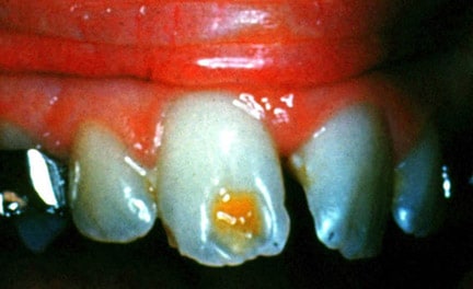
Thursday, September 9, 2010

Treatment of Crown Fractures With Pulp Exposure in Primary Incisors

Resident’s Name: Jessica Wilson
Program: Lutheran Medical Center - Providence

Article title: Treatment of Crown Fractures With Pulp Exposure in Primary Incisors

Author(s): Kupietzky, Holan.

Journal: Pediatric Dentistry

Year. Volume (number). Page #’s: 2003. 25(3). 241-247.

Major topic: Treatment of Crown Fractures

Overview of method of research: Clinical Review/Case Study

Purpose:
1. Present indications and contraindications for each type of treatment for complicated crown fractures in primary incisors.
2. Highlight advantages of partial pulpotomy (Cvek Pulpotomy) technique in certain scenarios.

Findings:
1. Partial Pulpotomy (PP)
a. Indications:
-Small, noncarious exposure occurring <14>14 days prior or extensive pulpal inflammation.

2. Cervical Pulpotomy
a. Indications:
-After considering PP, pulpal inflammation not extending past coronal portion with good hemostasis.
b. Technique:
- Formocresol: acute inflammation is fixed into chronic inflammation, roots do not continue to develop and teeth theoretically remain asymptomatic.
- Calcium Hydroxide (CH): teeth remain vital. Authors conclude that may be acceptable for mechanical exposures, but positive correlation between degree of inflammation and CH failure.
-If using CH for pulpotomy, absolute hemostasis is critical to prevent internal root resorption.
c. Contraindications:
-Infection or inflammation beyond coronal pulp with inability to achieve hemostasis as well as any PA pathology.

3. Pulpectomy
a. Indications:
-Trauma producing chronic inflammation or necrosis of radicular pulp and goal is to maintain esthetics and function.
b. Technique:
-Zinc Oxide Eugenol: not ideal due to resistance to resorption.
-Iodoform Paste: produce good results. Resorbable and producing antibacterial effects.
- CH: another possible option
c. Contraindications:
-Nonrestorable crown due to loss of tooth structure, extensive root resorption or PA infections extending to the permanent tooth bud.
-Parental concern about esthetics due to the high frequency of color change to yellow or brown.

4. Extraction:
-Considered as a last resort . Space maintenance generally not an issue after primary canine eruption. Discuss possibility of delayed or premature eruption of succedaneums dentition as well as possible effect on function, speech and appearance.

Key points/Summary:
1. PP technique may be used in immature or mature tooth, maintains tooth vitality, and provides esthetic result.
2. Additional studies on PP needed.

Assessment of Article: Clear & concise, but still need additional resources for an all inclusive review.

Intrusion Injuries of Primary Incisors. Part III: Effects on the Permanent Successors

Resident: Adam J. Bottrill
Date: 10SEP
Region: Providence
Article title: Intrusion Injuries of Primary Incisors. Part III: Effects on the Permanent Successors
Author(s):Diab, Mai DDS et al.
Journal: Quintessence International
Page #s: 377-384
Year: 2000:30
Major topic: Intrusion Injuries
Minor topic(s): NA
Type of Article: Analysis of Intrusion Injury Characteristics
Main Purpose: Analyze the effects of intrusion injury of primary incisors on the permanent dentision.

Key points in the article discussion:

I. General:

A. The potential for disturbances of the developing permanent dentition is high following injuries to their predecessors. (12-74% ???)

B. 18-69% (????) of permanent developmental defects due to INTRUSION injuries of primary teeth.

II. Factors Influencing the Sequelae of Intrusion Injuries.

A. Age of child:
1. Before 3yo, crown formation can be effected. (enamel hypoplasia, coronal dilaceration, odontoma etc...)
2. After 3yo, typically, root formation effected.
3. Typically, younger=more potential for permanent injury.
4. Germ can still suffer mineralization disturbances even after crown is fully formed.

B. Direction of intrusion:
1. Most risk when intrusion forces crown labially and root palatally.

C. Severity of Intrusion:
1. Increased severity = increased chance of damage
2. Alveolar fracture also increases chance.

D. Type of Treatment:
1. Several studies report that there is no correlation between severity of damage and the decision to extract versus allow to re-erupt.
2. Injury to the tooth is sustained at time of intrusion and not dependent on subsequent extraction.
3. The only exception to this rule is when subsequent infection occurs to intruded primary tooth.

III. Sequelae affecting coronal portions of the permanent successor:

A. White or yellow brown discoloration
1. Intrusion of primary teeth during mineralization of permanent crowns between 2-7yo.
2. Result of hypocalcified areas of the labial surface. (during "maturation" stage)
3. Bleeding may also hemoglobin products to enter the mineralizing portion of the enamel.

B. White of yellow-brown discoloration associated with enamel hypoplasia
1. "imperfect formation" due to injury between 2-3yo
2. displacement of the normal alignment of the ameloblast activity
3. irreversible destruction of the active enamel epithelium
4. grooves may form around the discoloration

C. Dilaceration of the crown:
1. Typically due to intrusion injury at around 2yo. (when 1/2 the crown is formed.
2. Displaced enamel epithelium becomes activated in a new/displaced position.
3. May erupt normally but will likely necrose... prophylactic crown recommended to avoid abscess.

IV. Sequelae Affecting Rot Portions of the Permanent Successors:

A. Duplication:
1. Rare malformation usually occurs from severe intrusion at around 2yo.

B. Dilaceration of the root:
1. Intrusion between 2-5yo.
2. Displaced hard tissue relative to the developing root... likely to be impacted.
3. Lateral dilaceration occurs between 2-7yo and usually tooth erupts normally.

C. Partial or complete cessation of root formation:
1. Rare sequelae due to intrusion between 4-7yo.
2. Hertwig's epithelial root sheath damaged.

V. Sequelae affecting the whole successor tooth:

A. Odontomalike malformation:
1. Severe intrusion of primary incisor between 1-3yo. (early stages of odontogenesis)
2. Require surgical extraction.

B. Sequestration of the permanent tooth germ:
1. Underdeveloped tooth germ and inadequate rot formation.
2. RARE... caused by SEVERE intrusion.
3. Also related to severe periradicular infection
4. Surgical extraction.

C. Disturbances of permanent successor eruption:
1. Early primary tooth loss (3-4yo) can cause delay in permanent successor eruption
2. Primary tooth loss later than 5yo can accelerate permanent eruption.
2. Ankylosed or delayed root resorption can also cause delayed eruption.

VI. Conclusion:

A. High probability of permanent tooth damage when primary tooth is intruded.
B. If the "wait and see" method is chosen... periodic recall is necessary to avoid periradicular infection and probably permanent tooth damage.

Assessment of article: Informative... BUT, the bottom line is, the permanent tooth is likely to be damaged. Knowing all the percentages of the type of sequelae doesn't really help me because it doesn't change treatment options. Also... despite the information presentd, I'm extracting the primary tooth if it's intruded. Not taking chances WRT subsequent infection.

Intrusion Injuries of Primary Incisors Part II: Sequelae affecting the intruded Primary Incisors


Department of Pediatric Dentistry
Resident’s Name: Murphy Program:Lutheran Medical Center - Providence
Article title: Intrusion Injuries of Primary Incisors Part II: Sequelae affecting the intruded Primary Incisors
Author(s): Mai Diab DDS MSc, Hossam Elbadrawy DDS MS
Journal: Quintessence International
Year. Volume (number). Page #’s: 2000. 31 #5. 335-341
Major topic: Intrusion Injuries and their follow up schedule/complications
Minor topic(s): Issues arising from intrusions
Main Purpose: Review Intrusion injuries
Overview of method of research: Clinical Review
Findings:
An intrusion injury occurs when a severe impact displaces the affected incisor deeper into the alveolar bone along the axis of the tooth causing trauma to the pulp and periodontal tissue. Intrusion injuries occur rather frequently in young children. Management of the injury depends on the extent of the intrusion and if there is an alveolar bone fracture. Radiographic exam must be completed to note the closeness of the developing tooth bud, alveolar bone fracture, and the amount of PDL that has diminished or eliminated.
The possible outcome of an intrusion injury is variable. The tooth may re-erupt with no complications, color changes may occur(gray, red, pinkish, bluish, black, or yellow), pulpal necrosis, resorption, or become anklosed.

Red/Gray Discoloration
Usually noticeable shortly after the injury. The gray color is caused by the traumatized pulp/tissue leaking heme into the dental tubules. This change in color may be reversible if the pulp survives the injury. Proper follow up is necessary. Some believe that a persistant gray color does NOT necessarily mean the pulp has necrosed, while others disagree. Frequent follow up appointments to monitor the tooth, and a good history from the patient will determine the necessary tx. If there is no pain or sensitivity, and no radiographic pathology, it’s ok to continue to monitor the tooth. If there is pain, etc, pulpectomy or EXT is indicated.

Yellow Teeth
This signifies calcification/obliteration of the pulpal canal. These should be treated the same as gray teeth, monitor it and treat any pertinent issues accordingly.

Necrosis
Loss of vitality is a common complication with intrusion injuries. The severity of the injury and the age of the child play in role in which way the pulp will react. More serious intrusions(bone fractures, >50% of the crown intruded) usually necrosis more often, and younger children (2-3) tend to show less necrosis than older children do. Tx options vary. Some suggest performing pulpectomies to try to save the tooth. Other disagree, stating that over instrumentation could cause injury to the developing tooth bud, and that the probability of ankylosis warrant EXT.

Abscess
Abscesses may occur with intruded teeth. If an abscess forms, EXT the tooth ASAP.

Ankylosis
Ankylosis is a real complication. Frequent recall exams to monitor if the tooth is in fact coming down is very important. If ankylosis is suspected with clinical exam(dull percussion, no reerupting), and a nonexistant PDL radiographically, take the tooth out ASAP.

Key points/Summary:
1. A proper exam at the time of injury is of great importance.
2. Frequent recall exams to assess how the tooth is doing must be done at 1-2 weeks, monthly for the first 3 months, and then every six months.
4. Monitoring the tooth is acceptable
5. Possible treatment includes doing nothing, performing a pulpectotmy, and EXT
6. Remember… the MOST important thing is the permanent tooth bud. If it’s in any danger, or may have been injured, try to get the primary tooth out ASAP.
Assessment of Article:
Great article. Every provider is going to differ on how they choose to treat intrusion injuries, and more often than not, they both may be correct.

09/10/2010 Complete intrusion of a maxillary right primary central incisor

Resident: J. Hencler
Date: 09/10/2010

Article title: Complete intrusion of a maxillary right primary central incisor
Author(s): Arthur Merkle, DMD

Journal: Pediatric Dentistry-22:2, 2000
Major topic: Trauma
Type of Article: Case Study

Main Purpose:
Present a rare case of complete intrusion of a maxillary right primary central incisor.

Findings:
A 29-month old female fell off a swing sustaining oro-facial injuries and was treated at the ED. Dentists on the hospital staff were not involved in the emergency care of this child. A plastic surgeon repaired oral and facial lacerations. A maxillary primary central incisor was assumed avulsed. Later, a routine dental exam of the child reveals an intrusion injury where the primary central incisor was displaced through the floor of the nasal cavity. IOE revealed a palpable mass in the maxillary labial vestibule near the anterior nasal spine. A mass was visible on the floor of the nasal cavity through the right naris. An occlusal radiograph confirmed the presence of the primary incisor and a lateral film revealed an intrusion injury in which the displacement was nearly one whole incisor length. Patient was referred to OS for EXT under GA. The intruded tooth was EXT through the right naris and had normal post –op recovery with uncomplicated healing.

Key points in the article discussion:
Luxation trauma is very common in the primary dentition. B/c alveolar bone in the young child in pliable, primary teeth are more likely to be luxated than fractured. Of luxation injuries, intrusive and extrusive traumas are the most common. If intruded sufficiently, the clinical presentation may suggest avulsion rather than intrusion. The apex of a completely intruded primary incisor will usually perforate the thin alveolar bone on the labial vestibule. This case is unusual b/c a maxillary right primary central incisor was intruded enough for the apex of the tooth to perforate the nasal cavity, so that avulsion was assumed.

Summary of conclusions:
This article emphasizes the importance of careful clinical and radiographic evaluation of dental trauma. Appropriate radiographs should be taken to verify clinical findings. In this case, earlier dx and tx would have placed the child and her developing dentition at less risk for possible complications associated w/ the intrusive injury.

Assessment of article:
Very interesting case. Take home message is this; “if you can’t see it, that doesn’t mean it’s not there.” Take a radiograph.

Friday, September 3, 2010

Traumatic Injuries in the Primary Dentition

Resident: Swan

Article Title: Traumatic Injuries in the Primary Dentition

Journal: Dental Traumatology

Volume (Number): 18; 2002, pgs 287-98

Major Topic: Injuries to the primary dentition

Type of Article: Literature Review

Main Purpose: update the “state of the art” regarding epidemiology and treatment of traumatic dental injuries in the primary dentition

Overview of method of research: Medline search and review of 75 articles that met inclusion criteria: clinical cases with no abstracts available and published before 1984 were excluded. Articles were sorted as pertaining to “epidemiology” or “therapy.”

Findings:

Epidemiology—

Prevalence of traumatic injuries in the 0-6 year old population varies from 11 to 30%. When the child starts walking alone between 18 and 30 months, the risk of trauma doubles compared to the average incidence for all children. In children up to age 2, avulsion and intrusion are the most severe injuries that can affect the developing tooth germ. Traumatic displacement of the root may alter the secretory phase of the ameloblast, leaving a defect known as circular enamel hypoplasia. A study of 255 traumatized primary teeth showed 23% with damage to corresponding permanent teeth. Highest incidence found after intrusion.

Therapy—Fractures

a) Non complicated crown fractures (in enamel and dentin)

a. PA taken as baseline, polish sharp edges, possibly restore with composite if child cooperates

b) Complicated crown fractures (w/ pulp exposure)

a. PA taken 1) partial pulpotomy if apex is open and child’s behavior allows it, 2) pulpotomy with formo/ZOE in cases where root is fully formed and not resorbing, 3) root canal treatment filled with ZOE, and 4) extraction

c) Crown-Root fractures (fracture compromises both crown and root)

a. PA, extract mobile fragments, leave root fragments to avoid permanent tooth germ damage

d) Root fracture

a. PA, 1) if coronal fragment is in place, root is complete, and pt cooperates, splint teeth. Tooth will have some mobility until the permanent tooth erupts, may lose crown 2) if crown is displaced (more common), extract coronal fragment

e) Alveolar fracture

a. PA/Pano, look for discontinuity in surrounding oral mucosa, reposition segment, splint for up to 4 weeks

Therapy—Luxations/Avulsions

a) Concussion (no mobility or sulcular bleeding)

a. Monitor, don’t do endo on discolored teeth (most are necrotic and asymptomatic)

b) Subluxation (mobile w/o displacement. May bleed)

a. Monitor. Most teeth regain stability within 2 weeks

c) Lateral luxation

a. With no occlusal interference, nearly all re-position spontaneously within a year

b. With interference, reposition and splint 2-3 weeks (reposition increases risk of necrosis

d) Intrusion

a. Apex is usually labially displaced (80%) On PA, apical tip can ben seen and tooth appears shorter than contralateral. If displaced toward permanent germ, apical tip is hidden and tooth appears elongated.

b. Palatally displaced—Extract, Labially displaced—leave for re-eruption (~88% re-erupt to occlusal plane)

e) Extrusion

a. Repostion and splint, or extract

Therapy—Avulsion

a) PA, do not replant

Instructions to Parents:

1) Soft diet for two weeks, 2) brush teeth after each meal, 3) chlorhexidine 2x daily for a week, 4) inform of possible complications and appearance of fistulae 5) w/ intrustion, avoid pacifier/bottle use to allow re-eruption

Key Points/Summary: Conservative treatment is the way to go with this population

Assessment of Article: Good overview of tx for primary teeth, research method not very statistically-based, hard to know how much weight to assign to their reported averages.

Intrusion injuries of primary incisors. Part I: Review and management

Department of Pediatric Dentistry

Resident’s Name: Jessica Wilson

Program: Lutheran Medical Center - Providence

Article title: Intrusion injuries of primary incisors. Part I: Review and management

Author(s): Diab, M et al.

Journal: Quintessence Intl

Year. Volume (number). Page #’s: 2000. 31(5). 327-334.

Major topic: Intrusion Injuries

Overview of method of research: Literature review

Findings:
Males have significantly higher incidence of primary tooth injuries after the first year.
Peak incidence of intrusion injuries between 1 and 3 years of age (root tips are fully formed) and rarely occurs after the age of 4 (root tips begin to resorb).
Primary incisors are highly susceptible as the alveolar bone contains large marrow spaces and high crown to root ratio.
Grade I (mild) intrusions: more than 50% of the crown is visible
Grade II (moderate) intrusions: less than 50% of the crown in visible
Grade III (severe) intrusions: complete intrusion of the crown
Although gingival bleeding and edema may be present, palpation and percussion sensitivity are rare.
Although one study on monkeys demonstrated there was less damage to permanent teeth if the intruded incisor is removed, other studies show there is no significant difference in frequency or
degree of damage to the permanent tooth.

Key points/Summary:
History of the accident as well as medical history is very important (antibiotics for endocarditis/tetanus immunization).
Diphtheria, pertussis, tetanus (DPT) vaccine given before 18 mo, then boosters given at 6 and every 10 years after. If child‘s injury has been exposed to soil and the child has not received vaccine in last 5 years, a booster is recommended.
It is not advised to separate young children from their parents at this time. Knee to knee and tell show do techniques may be indicated.
Neurological assessment is essential and if positive signs appear, hospitalization a must.
Extraoral and intraoral exams are to be performed. Although both upper and lower lip injuries are often present, contusions of the chin and lower lip are more prevalent.
Must take radiograph (PA & occlusal) to determine position orientation and integrity of intruded tooth as well as the alveolar bone surrounding it. The PDL is absent in the case of intrusion.
Labial crown inclination indicated palatal intrusion toward the unerupted permanent tooth. This appears elongated in the occlusal radiograph.
Less commonly seen, palatal crown inclination is indicative of labial intrusion away from the developing permanent tooth. This appears as a foreshortened root.
An anterolateral exposure may be indicated to determine the exact position of the intruded primary tooth. An occlusal film is taped to the child’s cheek, the x-ray beam is oriented from the opposite side and the exposure time is doubled.
Spontaneous re-eruption may occur within 1-6 months IF primary tooth is labially intruded or it is a class I intrusion. However, pulpal necrosis and pathologic root resorption may occur at a later time. Some authors recommend a 1 week dose of antibiotics to help prevent complications. If signs of re-eruption are not evident in 4-8 weeks, this may be indicative of ankylosis and extraction should be considered. Digit sucking may also be grounds for failure to re-erupt.
If signs or symptoms of infection at the site of trauma or pulpal infection/necrosis
All other circumstances such as a palatally intruded tooth, grade II or III intrusions, perforations of the buccal plate and alveolar bone fractures, extraction is indicated.
Whether or not to splint primary teeth is still controversial.
It is important to note that despite our best efforts to protect the developing permanent dentition, damage may still have occurred at the time of injury.
Recommendations to parents should include a soft diet, cleaning teeth with moist swab in alcohol-free mouthwash, and anticipating and signs or symptoms of infection in addition to follow up exams
Follow up appointments are indicated at 1 week post trauma, every 2 weeks for a month, then every 1 month for the next three months and finally every 6 months. A follow-up radiograph should be taken 1-2 months post trauma to verify proper healing.

Assessment of Article: Great summary of recommendations and the research backing them.

Thursday, September 2, 2010

Developmental disturbances of the permanent teeth following trauma to the primary dentition

Meghan Sullivan Walsh September 2, 2010

Literature Review - LMC/ Providence Pediatric Dentistry


Developmental disturbances of permanent teeth following trauma to the primary dentition.


Resident: Meghan Sullivan Walsh


Program: Lutheran Medical Center - Providence


Article Title: Developmental disturbances of permanent teeth following trauma to the primary dentition.


Authors: Thomas von Arx, DMD


Journal: Australian Dental Journal


Volume Number, Year, Pages ; 38:1 1993, Pages 1-10


Major Topic: Study of permanent tooth malformation following trauma to the primary dentition.


Overview of Method of Research: One hundred and ninety-five children treated for a dental emergency in the Department of Oral and Maxillofacial Surgery, Kantonsspital, Lucerne, Switzerland between 1977 and 1998 were recalled in 1990. Of these children one hundred and fourteen children could be reexamined. The children were categorized into groups based on original trauma, eruption stage of permanent succedaneous tooth and developmental disturbances to the permanent teeth.


Key Points: The total number of traumatized teeth in these patients were 255. The majority of injuries affected the maxillary central incisors then laterals. Male to female ratio was 1.6:1 and the mean age at time of trauma was 3.6 years old. The study showed that 23 percent of the reexamined teeth showed developmental disturbances. Approximately one in four teeth interfered with the odontogenesis of it’s successor. The majority of tooth disturbances were enamel hypoplasia and or enamel defects. Intrusion bore the highest risk of tooth disturbances with 54% of these injuries resulting in some affect of the permanent dentition. No disturbance was found in any of the cases with a crown of root fracture even when the pulp was exposed. All the other types of developmental disturbances were seen to have some correlation to time at which the primary tooth was injured. Odontome-like teeth were most likely to develop following injury at an early age such as one year old. Crown dilaceration occurred mostly in the age range of 1.5-3.5 and root malformation occurred later at four and five years of age. The author concluded that enamel continues to maturate and mineralize until the time of eruption which can explain why enamel disturbances and discoloration may effect all age groups.


Assessment of the Article: This was a great clean and simple study. I was impressed with the number of patients they were able to follow up with especially many years after sustaining the injury. The author did note that his numbers may be off due to the fact that only 56% of these teeth could be examined clinically. Many of these patient’s did not have their permanent teeth yet erupted and so these teeth were examined radiographically. We should make note of the high incidence of damage to the permanent dentition especially following an intrusion and remember to tell our parents that is is likely that their permanent dentition will be effected.

Trauma!


Leforte Fractures


Intraoral electrical burn
Department of Pediatric Dentistry
Resident’s Name: Murphy Program:Lutheran Medical Center - Providence

Article title: Trauma, Chapter 9
Author(s): numerous
Journal: The Handbook of Pediatric Dentistry
Year. Volume (number). Page #’s:
Major topic: Trauma
Minor topic(s): Trauma assessment from beginning to end
Main Purpose: List all the necessary/recommended steps in the process of working up a child who has had a traumatic injury
Overview of method of research: Review

Findings:
This ‘article’ was simply Chapter 9 out of “The Handbook”. It’s already been broken down, so it’s kind of hard to summarize. I will attempt it anyway. First triage the situation.
Is the patient conscious? Are they vomiting, nauseas, seizing, etc.
Is the patient responsive? Can they tell you what happened?
Who else was there at the time of the incident
Use the Glasgow coma scale for eval. Of head trauma.

A thorough exam should include a thorough medical/dental history, notes on what/when/where/how the incident happened, extra/intra oral exam, and any necessary x-rays should be taken. After all of the necessary information has been gathered, a diagnosis should be given with a proposed treatment, and then treatment should be initiated. Once completed, any necessary meds should be prescribed, and there should be a follow up plan.
Possible trauma situations include but are not limited to fractured, displaced, or missing teeth, pulp exposures, bone fractures, lacerations, degloving, electrical burns, etc.
The Handbook has a great 5 page flow chart that is included in your residence manual. Definitely a great idea to bring it with you to your ED calls.
Remember we do NOT reimplant primary teeth due to ankylosis, infections risk, and possibly injuring the perm. Tooth bud.

Possible complications
Color changes (gray). Tx may not be indicated, sometimes the tooth changes back. Monitor it routinely.
Pulpal obliteration(calcification), necrosis, internal/external resorption.
Damage to developing tooth bud
ankylosis


Key points/Summary:
The second years last year said there was a lot of questions on the boards with regards to lacerations and electrical burns.

Soft Tissue Injuries
Tongue Lac-suture if bleeding is not controllable
Through and through punctures-suture both sides after adequate debridement
Vermillion border lacs-suture starting EXTRA orally
Ensure proper debridement!


Electrical Burns
Deep burns 2000 C- usually painless
eschar sloughs 7-10 days/bleeding from facial artery possible
use an appliance to stop contraction of the wound, worn for 6-12 months, usually with topical antibiotics
Plastic surgery usually necessary

Assessment of Article: Sort of tough to summarize a summary. We all should know The Handbook backwards and forwards…. So check it out!

09/02/2010 Guidelines for the management of traumatic dental injuries. III. Primary Teeth

Resident: J. Hencler
Date: 09/02/2010

Article title: Guidelines for the management of traumatic dental injuries. III. Primary Teeth

Author(s): Flores et al.
Journal: Dental Traumatology 2007; 23: 196-202

Major topic: Trauma guidelines in primary teeth

Overview of method of research:
The IADT has developed guidelines that represent the current best evidence based on literature research and professional opinion.

Guidelines:

Uncomplicated Crown Fracture: Pulp not exposed. Tx: Smooth sharp edges-tooth restored w/ comp.

Complicated Crown Fracture: Pulp exposure. Tx: Preserve pulp vitality by pulp capping or partial pulpotomy. CaOH is a suitable material.

Crown-root fracture: Involved enamel, dentin, and root structure. May or may not involve pulp. Tx: EXT

Root fracture: coronal fragment is mobile. Tx: EXT coronal fragment, apical fragment left to resorb.

Alveolar Fracture: Tooth containing segment often mobile and usually displaced. Tx: reposition and splint.

Concussion: tooth is painful to touch, not mobile. Tx: observation.

Subluxation: Mobility noted but not displaced, bleeding from gingival sulcus may be noted. Tx: observation

Extrusive Luxation: Tooth appears elongated and is mobile. Tx: based on degree of displacement. For minor extrusion <3mm reposition or allow for spontaneous reposition. Severe extrusion=EXT.

Lateral luxation: Tooth is displaced and mobile. Tx: If no occlusal interference, allow tooth to spontaneously reposition. If occlusal interference observed, give LA and reposition. Severe displacement=EXT.

Intrusive luxation: displaced through labial bone or impinging on succedaneous tooth bud. Tx: If apex is displaced through labial bone plate, let tooth spontaneously reposition. If apex displaced into developing tooth bud=EXT.

Avulsion: Tooth out of socket. Tx: Do not replant tooth!

Discussion:
It is important to take a thorough med hx and hx of present illness. After clinical exam, radiographs should be considered depending on child’s ability to cope w/ procedure. Depending on child’s injury, typical radiograph may include a 90° horizontal-view, occlusal view, or extra-oral lateral view (useful to reveal relationship b/t apex and perm tooth bud). Post op patient instruction should include recommendations such as OHI and diet. Chlorhexideine (0.1%) applied topically to the affected area is often beneficial 2x/day for 2 wks. Inform parents of future complications such as defects in perm tooth structure or possible loss of traumatized primary tooth. Finally, give appropriate f/u appointment for post-op care and re-evaluation. (See last table)

Summary of conclusions:
An appropriate tx plan after an injury is important for a good prognosis. Guidelines are useful for dentists and other healthcare professionals in delivering the best care possible in an efficient manner. Management of trauma to primary teeth differs from that used in permanent teeth. Treatment selection should minimize any risk of further damage to permanent successor.

Assessment of article:
This is a great article that provides very useful clinical information that all pediatric dentists should know and/or can refer to when treating trauma cases. These guidelines combined with sound clinical judgment make treating traumatic injuries to primary teeth straightforward and more predictable.