Saturday, April 30, 2011

Trico-dento-osseous Syndrome


Definition: Tricho-dento-osseous (TDO) syndrome is an autosomal dominant genetic disorder that belongs to the group of diseases known as ectodermal dysplasias. It derives its name from the three primarily affected tissues including hair, teeth and bone.

Etiology: It is caused by a DLX3 gene mutation. Research suggests that Amelogenesis imperfecta of the hypomaturation-hypoplasia type with taurodontism and TDO are two genetically distinct conditions.

Clinical Features: TDO syndrome is characterized by kinky or curly hair; poorly developed tooth enamel; and unusual thickness and/or denseness (sclerosis) of the top portion of the skull (calvaria) and/or the long bones. In some cases, affected individuals also exhibit abnormally thin, brittle nails or premature fusion of the fibrous joints between certain bones in the skull (craniosynostosis), causing dolicocephaly.
Kinky, course and/or curly hair is present at birth in 80% of people with TDO. Only 46% of the individuals with TDO and kinky, course and/or curly hair at birth retain this phenotype after infancy. There is an increased cranial thickness. The loss of visible mastoid pneumatization is the most common osseous feature seen in affected individuals (82%) and is relatively uncommon in unaffected people (8%).

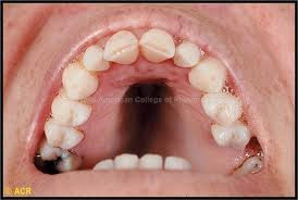
Oral Manifestations: While all individuals with TDO appear to have enamel hypoplasia and taurodontism, the expression of these traits is highly variable. The teeth appear discolored in 76% of the affected individuals while the remaining affected individuals have teeth of normal color. Enamel alteration in people with TDO ranges from being extremely thin and/or rough and pitted to being of normal color and only slightly decreased thickness.

Treatment: Symptomatic.

Dental Considerations: Treatment depends on severity of defects and esthetic demands of patient and may include full coverage restorations.

Lesch-Nyhan Syndrome


Definition: Lesch-Nyhan syndrome is an inheritable disorder that affects how the body builds and breaks down purines.


Etiology: inherited as an X-linked trait. It mostly occurs in boys. Persons with this syndrome are missing or are severely lacking an enzyme called hypoxanthine guanine phosphoribosyltransferase 1 (HGP). The body needs this enzyme to recycle purines. Without it, abnormally high levels of uric acid build up in the body.


Frequency: about 1 in 380,000 people.

Clinical Features: The excess uric acid levels cause children to develop gout-like swelling in some of their joints. In some cases, kidney and bladder stones develop because of the high uric acid levels. Males with Lesch-Nyhan have delayed motor development followed by bizarre, sinuous movements and increased deep tendon reflexes. A striking feature of Lesch-Nyhan syndrome is self-destructive behavior characterized by chewing off fingertips and lips, if not restrained that begins in the second year of life. It is unknown how the enzyme deficiency causes these problems.

Oral Manifestations: The most typical feature results in partial or total destruction of perioral tissues.


Treatment: Treatment for LNS is symptomatic. Gout can be treated with allopurinol to control excessive amounts of uric acid. Kidney stones may be treated with lithotripsy, a technique for breaking up kidney stones using shock waves or laser beams. There is no standard treatment for the neurological symptoms of LNS. Some may be relieved with the drugs carbidopa/levodopa (used to treat Parkinson's disease), diazepam, phenobarbital, or haloperidol (antipsychotic).

Dental Considerations: A soft mouthguard fabricated to prevent the destruction of perioral soft tissues and combined psychiatric pharmacologic therapy proved to have satisfactory results. In extremem cases, teeth may need to be extracted.

Prognosis: The outcome is likely to be poor. Persons with this syndrome usually require assistance walking and sitting and generally need a wheelchair to get around. Death is usually due to renal failure in the first or second decade of life.

Treacher Collins Syndrome


Treacher-Collins syndrome is a genetic condition characterized by a defective protein called treacle, which affects the development of bones and other tissues in the face. The signs and symptoms of this disorder vary greatly, ranging from almost unnoticeable to severe. Over 50% of cases are thought to be caused by a new genetic mutation as there is no family history.


Symptoms:
· Outer part of the ears are abnormal or almost completely missing
· Hearing loss
· Very small jaw (micrognathia)
· Very large mouth
· Defect in the lower eyelid (coloboma)
· Scalp hair that reaches to the cheeks
· Cleft palate


Signs and tests: The child usually will show normal intelligence. Genetic tests can be done to look for mutations in the TCS1 gene. Examination of the infant may reveal a variety of problems, including:
· Abnormal eye shape
· Flat cheekbones
· Clefts in the face
· Small jaw
· Low-set ears
· Abnormally formed ears
· Abnormal ear canal
· Hearing loss


· Defects in the eye (coloboma that extends into the lower lid)
· Decreased eyelashes on the lower eyelid


Treatment: Treatment involves testing for and treating any hearing loss so that a child can perform at a normal level in school. Plastic surgery can treat the receding chin and other defects.


Prognosis: Children with this syndrome typically grow to become normally functioning adults of normal intelligence. Careful attention to any hearing problems helps ensure better performance in school.Complications: Feeding difficulty, speaking difficulty, communication problems, vision problems.

Friday, April 29, 2011

Sturge-Weber Syndrome


Definition: Sturge-Weber Syndrome (encephelotrigeminal angiomatosis) is a congenital, non-familial hamartomatous vescular proliferation mainly involving areas along the trigeminal nerve distribution.

Etiology: Not clear, but is thought to be caused by the presence of a vascular plexus around the portion of the neural tube destined to become the facial skin.

Frequency: Rare.

Clinical Features: Hemangiomas of the skin, face, and oral mucosa, calcifications of the brain, ocular disorders such as glaucoma, epilepsy and mild mental retardation. Facial hemangiomas are the most constant and characteristic finding of this syndrome appear as a bright red or purple and are usually unilateral.


Oral Manifestations: Oral hemangiomas also tend to be unilateral, may involve the maxillary gingival, buccal mucosa, tongue and lips, are usually flat, but may have a raised irregular surface that causes tissue enlargement. Delayed or early and ectopic dental eruptions are also common.

Treatment: Depends on the location and severity of the lesions, laser therapy may lighten or remove stains. Anticonvulsants may be indicated.


Dental Considerations: Symptomatic.

Neurofibromatosis


The neurofibromatoses are genetic disorders that cause tumors to grow in the nervous system. The tumors begin in the supporting cells that make up the nerves and the myelin sheath. These disorders cause tumors to grow on nerves and produce other abnormalities such as skin changes and bone deformities. Although many affected persons inherit the disorder, between 30 and 50 percent of new cases arise spontaneously through mutation in an individual's genes. Once this change has taken place, the mutant gene can be passed on to succeeding generations.

Neurofibromatosis type 1 (NF1) is the most common type of the neurofibromatoses. Changes in skin appearance, tumors, or bone abnormalities occur. Symptoms of NF1, which may be evident at birth and nearly always by the time the child is 10 years old, may include light brown spots on the skin "cafe-au-lait" spots), two or more growths on the iris of the eye, a tumor on the optic nerve, a larger than normal head circumference, and abnormal development of the spine, a skull bone, or the tibia.
Treatment: Surgery is often recommended to remove the tumors. Some NF1 tumors may become cancerous, and treatment may include surgery, radiation, or chemotherapy.
Prognosis: In most cases, symptoms of NF1 are mild, and individuals live normal and productive lives. In some cases, however, NF1 can be severely debilitating and may cause cosmetic and psychological issues.

NF2 is less common and is characterized by slow-growing tumors on the eighth cranial nerves. The tumors cause pressure damage to neighboring nerves. Tumors in the 8th nerve, cataracts at an early age or changes in the retina that may affect vision, other nervous system tumors are manifestations. It often starts in the teen years.
Treatment: MRIs can reveal tumors as small as a few millimeters in diameter, thus allowing early treatment. Surgery to remove tumors completely is one option but may result in hearing loss.
Prognosis: The course of NF2 varies greatly among individuals. In some cases of NF2, the damage to nearby vital structures, such as other cranial nerves and the brain stem, can be life-threatening.

Schwannomatosis is the rarest of the three types and is characterized by the development of multiple schwannomas everywhere in the body except on the vestibular branch of the 8th cranial nerve. The dominant symptom is pain, which develops as a schwannoma enlarges or compresses nerves or adjacent tissue. Some people may develop numbness, tingling, or weakness in the fingers and toes.
Treatment: There is no currently accepted medical treatment or drug for schwanomatosis, but surgical management is often effective. Pain usually subsides when tumors are removed completely.
Prognosis: Most individuals with schwannomatosis have significant pain. In some extreme cases the pain will be severe and disabling.

Oral lesions occur in 60-70% of cases and are characterized by multiple or isolated nodular neurofibromas, which vary in size. Tumors usually involve the tongue although other areas of mucosa may be affected. Enlargement of the fungiform papilla is common. Macroglossia is less common and lesions of the maxilla and mandible are relatively uncommon.

Marfan Syndrome


Definition: An autosomal dominant disorder of the connective tissue. MFS features can occur in many different parts of the body.


Etiology: Defects in a gene called fibrillin-1. Majority of the cases are genetic, however, up to 30% of cases have no family history.
Frequency: About 1 in 5,000.

Clinical Features: Long, thin arms and legs, scoliosis and arachnodactyly. Arm span is much greater than their height. A chest that sinks in or sticks out -- funnel chest or pigeon breast. Flat feet, hypotonia, hyperflexible joints, learning disabilities, severe myopia or dislocation of the lens of the eye, aortic dilation or aortic aneurysm, heart valve problems, or collapsed lung.


Oral Manifestations: Long, narrow face, high palatal vault, prominent lower jaw, crowding and malocclusion.

Treatment: Vision problems should be treated when possible. Treatment of scoliosis in adolescence, medication to slow the heart rate may help prevent stress on the aorta, avoiding participating in competitive and contact sports to avoid injuring the heart. Some people may need surgical replacement of the aortic root and valve.

Dental Considerations: Antibiotic prophylaxis and orthodontic care.
Prognosis: Heart-related complications may shorten the lifespan of people with this disease. However, many patients survive well into their 60s. Good care and surgery may extend the lifespan further.

Wednesday, April 27, 2011

Beckwith-Wiedemann Syndrome

Syndrome: Beckwith Wiedemann Syndrome
Etiology: Congenital overgrowth disorder that is caused by a sporadic genetic mutation in 85% of cases. Genetics are complex, but chromosome 11 has been implicated as the problem area. BWS remains a clinical diagnosis because physicians cannot identify and test for all the genetic causes of BWS.
Five common features used to define BWS are: macroglossia (large tongue), macrosomia (birth weight and length >90th percentile), midline abdominal wall defects (omphalocele-intestines, liver, other organs remain outside the abdomen in a sac b/c of defect in abdominal wall muscles, umbilical hernia, diastasis recti-separation of rectus abdominis muscles into right/left halves), ear creases or ear pits, and neonatal hypoglycemia (low blood sugar after birth).
Diagnosis: Patients normally don’t present with all 5 features. Child is considered to have the syndrome if it has been diagnosed by a physician and they present with at least 2 of the 5 major features.
Systemic/Medical Conditions: Patients have higher risk of developing cancer, although 80% do not. Most common tumors are Wilms’ tumor (nephroblastoma) and hepatoblastoma. Both can usually be cured if diagnosed early.
Oral Manifestations: Macroglossia (one of the five principal features)
Dental needs/considerations: Macroglossia in BWS becomes less noticeable with age and often requires no treatment; but it does cause problems for some patients. In severe cases, macroglossia can cause respiratory, feeding, and speech difficulties. Children with BWS and significant macroglossia should be evaluated by a craniofacial team. Early orthodontic intervention can halt problems before they progress (open bite, mouth breathing, protrusion of teeth)
The best time to perform surgery for a large tongue is not known. Some surgeons recommend performing the surgery between 3 and 6 months of age. Surgery for macroglossia involves removing a small part of the tongue so that it fits within the mouth to allow for proper jaw and tooth development.

de Lange Syndrome

Syndrome: De Lange Syndrome (Cornelia de Lange Syndrome)
Etiology: Genetic disorder that can lead to serious developmental anomalies, affecting both physical and intellectual development of a child. Most due to spontaneous mutations. No specific gene associated.
Diagnosis: solely a clinical diagnosis right now. There are no biochemical or chromosomal markers for DLS yet.
Systemic/Medical Conditions:
• Low birth weight (usually under 5 pounds / 2.5 kilograms)
• Delayed growth and small stature
• Developmental delay
• Limb differences (missing limbs or portions of limbs)
• Small head size (microcephaly)
• Thick eyebrows, which typically meet at midline (synophrys)
• Long eyelashes
• Short upturned nose and thin downturned lips
• Long philtrum
• Excessive body hair
• Small hands and feet
• Small widely spaced teeth
• Low-set ears
• Hearing impairments
• Vision abnormalities (e.g., ptosis, nystagmus, high myopia, hypertropia)
• Partial joining of the second and third toes
• Incurved 5th fingers
• Gastroesophageal reflux
• Seizures
• Heart defects
• Cleft palate
• Feeding problems
Children with this syndrome are often found to have long eyelashes, bushy eyebrows and synophrys- Unibrow!
Children with CdLS often suffer from gastrointestinal tract difficulties, particularly gastroesophageal reflux. Vomiting, intermittent poor appetite, constipation, diarrhea or gaseous distention are known to be a regularity in cases where the GE tract problems are acute. However, symptoms may range from mild to severe.
Oral Manifestations: Some dental abnormalities reported include delayed eruption, spacing and macro- or microdontia.
Recent Info: 2009 study out of Turkey found possible correlation between CdLS and Hutchinson’s teeth.

Monday, April 25, 2011

Trisomy 9 Mosaicism


Definition: 9th chromosome appears three times (trisomy) rather than twice in some cells of the body. The term "mosaic" indicates that some cells contain the extra chromosome 9, while others have the normal chromosomal pair.

Etiology: Errors during meiosis or mitosis

Frequency: Rare

Diagnosis: Take samples from multiple tissues for diagnosis

Clinical features: Associated symptoms and findings may vary greatly in range and severity, depending on the percentage of cells with the extra chromosome.

Growth deficiency before birth, mental retardation, congenital heart defects, craniofacial abnormalities (sloping forehead, bulbous nose, short palpebral fissures, deeply set eyes, and low-set ears, small jaw, large fontanels), musculoskeletal, genital, kidney issues.

Rubenstein-Taybi Syndrome


Other names: Broad thumb-hallux syndrome or Rubinstein syndrome

Definition: Condition characterized by short stature, moderate to severe learning difficulties, distinctive facial features, and broad thumb and first toes.

Etiology: Autosomal Dominant

Frequency: Uncommon 1:125,000

Clinical features: Broad thumbs and broad first toes, mental disability, small height, bone growth, small head, cryptorchidism in males (absence of one or both testes from the scrotum), unusual facial features (flat red birthmark on forehead, widely spaced eyes, downward slant of eyes, strabismus (cross eyed), droopy eyelids, high arched eyebrows, beaked nose, broad nasal bridge, extra fold of skin on either side of nose, malformed ears, high-arched palate, crowded teeth, small lower jaw), hirsutism (excess hair on body), hyperextensible joints, small tilted pelvis, seizures, slow development of cognitive and motor skills along with low muscle function, unsteady gait, feeding difficulties, respiratory infections, eye infections, heart defects, vertebral abnormalities, G.I. reflux, and kidney problems

Increased risk of noncancerous and cancerous tumors, leukemia, and lymphoma

Treatment: Individuals with RTS have a range of mild to severe symptoms. Most children will benefit from speech therapy. Some children may not be able to speak verbally, and therefore, would may need to learn sign language.

Cri-du-chat


Cri-du-chat: Group of symptoms that result from missing piece of chromosome number 5. Syndrome based on the infant’s cry, which is high-pitched and sounds like a cat.

Etiology: Genetic information on chromosome 5 is missing. One missing piece is called TERT (telomerase reverse transcriptase).

Diagnosis: Made at birth by clinical observation. Infant’s cry sounds like a high-pitched cat.

System/medical conditions associated: Cry that is high-pitched and sounds like a cat. Downward slant to the eyes. Low birth weight and slow growth, low-set or abnormally shaped ears, mental disability, partial webbing or fusing of fingers or toes, single line in the palm of the hand, skin tags, slow or incomplete development of motor skills, small head, small jaw, wide set eyes. Cardiac abnormalities which may require surgical correction.

Oral manifestations: Small jaw, speech delay. Cleft lip and palate.

Dental needs: Frequent examination and prevention techniques. Treatment of special needs patient. Cleft lip and palate treatment.

Recent information: Children may be treated by speech, sound and occupational therapists.

Ehler Danlos Syndrome


Ehler Danlos Syndrome: Group of inherited connective tissue disorders caused by a defect in synthesis of collage.

Etiology: Mutation in the following – Fibrous proteins: COL1A1, COL1A2, COL3A1, COL5A1, COL5A2 and TNXB or Enzymes: ADAMTS2, PLOD1. Mutations in these genes alter the process or collagen or proteins that interact with collagen.

Diagnosis: Made through clinical observation. Skin biopsy is useful.

System/medical conditions associated: highly flexible fingers and toes, loose unstable joints, flat feet, joint pain without inflammation, debilitating fatigue, high and narrow palate, vulnerability to chest and sinus infections, easy bruising, fragile blood vessels, smooth, stretchy skin, abnormal wound healing, low muscle tone, early onset of osteoarthritis, cardiac effects such as valvular heart disease, difficulty regulating blood temperature, severe mouth ulcers, food allergies, sensitivity to medication, insensitivity to local anesthetics, migraines and headaches, fibromyalgia, arachnodactyly. Less common are osteopenia, deformities of the spine, functional bowel disease, nerve compression disorders, vascular skin conditions, blue sclera, otosclerosis, premature rupture of membranes, platelet aggregation failure, swan neck deformity of the fingers.

Oral manifestations: Abnormal wound healing, thin and fragile mucosa, easy bleeding and bruising. Patients can touch the tip of their nose with their tongue. Enamel, dentine and cementum defects. Increased tendency to develop multiple pulp stones and TMJ hypermobilty.

Dental needs: Prevention of dental disease, periodontitis. Awareness of insensitivity to anesthetics.

Recent information: Outlooks for EDS depends of the severity of disease. Lifelong disease, no cure.

Wednesday, April 20, 2011

William's Syndrome


Williams Syndrome: Rare genetic development that can lead to developmental problems


Etiology:Rare genetic disorder caused by missing genes. A person with Williams syndrome has a 50% chance of passing on the syndrome to their children. Occurs in 1:8,000 births.


Diagnosis: Diagnosis not always made at birth. Signs and symptoms can lead to blood work and genetic testing to determine diagnosis.


Systemic/medical conditions associated: Delayed speech that may later turn in strong speaking abilities. Developmental delays, easily distracted, ADD, feeding problems, clinodactyly or inward bend of the small finger, learning disorders, and mild to moderate mental disabilities. Personality traits include being very friendly, trusting strangers and interested in music. Short stature and sunken chest. Medical conditions include aortic stenosis, pulmonary stenosis. High blood pressure and hypercalcemia which may cause seizures.


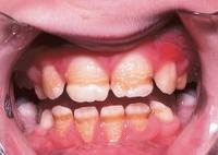
Oral manifestations: Facial manifestations: Flatterned nasal bridge with upturned nose, long riges in skin, prominent lips with open mouth, epicanthal folds, partially missing teeth, defective tooth enamel or small widely spaced teeth.


Dental needs: Early treatment and prevention for those with defective tooth enamel. Making patient comfortable, non stress inducing appointment due to poor heart condition and blood pressure. General Anesthesia is more risky in these patients because of these conditions.

Recent information: Avoid taking extra calcium and vitamin D. Physical therapy and speech therapy is important for these patients.

Hurler Syndrome


Hurler Syndrome: Also known as muciopolysaccharidosis. Genetic disorder that results in the buildup of glycosaminoglycans.


Etiology: Results in the buildup of glycosaminoglycans due to a deficiency of alpha-L iduronidase. A buildup of heparan sulfate and dermatan sulfate occurs. Occurs in 1:25,000 births.


Diagnosis: Prenatal diagnosis with amniocentesis and chorionic villus sampling can verify if a fetus either carries a copy of the defective gene or is affected with the disorder. After birth of a child clinical examination and urine tests are used.


Systemic/medical conditions associated: Progressive deterioration, hepatosplenomegaly, dwarfism and unique facial features. Mental retardation is progressive with death occurring by age 10. Language may be limited due to hearing loss and enlarged tongue. Corneas become clouded and retinas begin to degenerate. Carpal tunnel syndrome and restricted joint movement is common. Children may be large at birth with hernias. Many children slow in growth after age three develop a short body trunk and grow to less than 4 feet. Facial features include flat face, depressed nasal bridge and bulging forehead. Liver spleen and heart are often enlarged. Feeding often difficult and many children die early from obstructive airway disease, respiratory infections or cardiac complications.


Oral manifestations: Facial manifestations: Monitor for obstructive airway when performing dental treatment.


Dental needs: Prevention and often and early treatments to prevent stressful and lengthy appointments.


Recent information: Gene therapy has gained much interest. Patients are given retroviral, lentiviral, AAV and even nonviral vectors to deliver the iduronidase gene. This treatment has been successful on mice, dogs and cat models.

Tuesday, April 19, 2011

Goldenhar Syndrome


Resident: Swan
Syndrome: Goldenhar Syndrome, AKA Oculo-Auriculo-Vertebral (OAV) syndrome
Etiology: congenital defect characterized by incomplete development of the ear, nose, soft palate, lip, and mandible, usually unilateral. Often synonymous with hemifacial microsomia. It is associated with anomalous development of the first and second branchial arches
Diagnosis: No genetic test available. Diagnosis made by physician after evaluation of characteristic symptoms.
Systemic/Medical Conditions: Some patients will have growing problems with various organs, especially heart, kidney, lungs. Usually, the organ will either not be present on one side or will be underdeveloped. Also, possible scoliosis, hearing loss, limbal dermoids (benign congenital tumor of the eye, where cornea meets sclera), strabismus is common, abnormal ear formation, tissue tags near the ear.
Oral Manifestations: hypoplastic mandible/maxilla, possible cleft lip/palate, facial asymmetry, high vaulted palate, class III malocclusions, anterior open bite.
Dental needs/considerations: possible distraction osteogenesis with bone grafting necessary

Achondroplasia


Resident: Swan
Syndrome: Achondroplasia

Etiology: Sporadic mutation in 85% of cases (2 parents without achondroplasia may give birth to child with it), or autosomal dominant pattern of inheritance. It is an abnormality of cartilage formation brought on by a change in the DNA for fibroblast growth factor receptor 3. It’s the most common type of dwarfism. If a homozygous child receives copies of gene from both parents, very unlikely the child will survive more than a few months.

Diagnosis: Prenatal ultrasound may show excessive amniotic fluid surrounding the unborn infant. After birth, the infant shows increased front to back head size, along with possible signs of hydrocephalus. X-rays of the long bones can reveal the condition in the newborn.

Signs/Symptoms: Characteristic space between the long and ring fingers (“trident hands”), bowed legs, decreased muscle tone, relatively large head size, prominent forehead, short upper arm and thigh, short stature, spinal stenosis

Oral Manifestations:

Craniofacial: Macrocephaly, prominent forehead (bossing), depressed nasal bridge, maxillary hypoplasia, lip incompetence, foramen magnum stenosis. These may lead to hydrocephalus, apnea, upper-airway obstruction, otitis media, sinusitis, and dental malocclusion.

Dental: Anterior open bite, Class III malocclusions (due to maxillary hypoplasia), possible delayed dental development

Dental needs/considerations: Early Ortho evaluation/intervception for Cl. III malocclusion, place a cushion behind child’s back to reduce back pain (spinal stenosis).

Sunday, April 17, 2011

Klinefelter Syndrome


Definition: condition in which human males have an extra X chromosome “XXY syndrome

Etiology: Nondisjunction. Nondisjunction occurs with when homologous chromosomes fail to separate.

Frequency: 1:1000

Clinical features:

- Tall height

- Abnormal body proportions (long legs, short trunk, shoulder equal to hip size)

- Small testicles

- Reduced fertility

- Abnormally large breasts (gynecomastia)

- Less than normal amount of facial and pubic hair

- Oral: taurodontism (elongated pulp chamber with short stunted roots)

Diagnosis:

- May first be diagnosed when a man seeks a physician consult due to infertility

Treatment:

- Testosterone therapy may aid in producing a more masculine appearance.

Complications:

Increased risk of:

- ADHD

- Autoimmune disorders such as SLE, Rheumatoid Arthritis, Sjogren Syndrome

- Breast cancer in men

- Depression

- Learning disabilities, including dyslexia

- Extragonadal germ cell tumor

- Lung disease

- Osteoporosis

- Varicose Veins

Turner Syndrome


Definition: Genetic condition in which a female does not have the usual pair of two X chromosomes. The condition only occurs in females. Most commonly, the female patient has only one X chromosome – “monosomy X”. Others may have two X chromosomes, but one of them is incomplete. In some cases, the chromosome is missing in some cells but not others “mosaicism”.

Etiology: Genetic mosaicism (46XX/45XO), nondisjunction (45XO), and partial monosomy (46XX). During conception, part of all of the second sex chromosome is not transferred to the fetus.

Frequency: 1:2000

Clinical features:

Possible symptoms in young infants:

- Lymphedema: swollen hands and feet

- wide and webbed neck

Possible general symptoms:

- short height

- increased weight, obesity

- absent or incomplete development at puberty

o sparse pubic hair and small breasts

- broad, flat chest shaped like a shield “shield chest”

- drooping eyelids

- dry eyes

- low hairline

- low-set ears

- infertility

- absent menstruation

- vaginal dryness, can lead to painful intercourse

Oral features:

- micrognathia: small lower jaw

- high-arched palate

Treatment:

Short height -> Growth Hormone

Lack of development of sexual characteristics -> Estrogen

Infertility -> Donor Egg

Concurrent Complications:

- congenital heart disease and other cardiac problems

- horseshoe kidney

- hypothyroidism

- diabetes

- vision problems

- hearing problems

- autoimmune diseases

- ADHD

- cognitive deficits (visuospatial, mathematical, and memory areas)

Tuesday, April 12, 2011

Crouzon Syndrome


Syndrome: Crouzon Sydrome (Craniofacial Dysostosis)
Etiology: Autosomal dominant inheritance, with complete penetrance and variable expressivity. Missense mutation in FGFR2 gene. Results when premature fusion of cranial sutures occurs. This initiates changes in the brain secondary to increased intracranial pressure. Deformities of cranial bones and orbital cavities also result.
Diagnosis: prenatal testing can diagnose exophthalmos, which can foreshadow this type of developmental problem. However, the phenotypic features of Crouzon syndrome may be absent at birth and evolve gradually during first few years of life.
Systemic/Medical Conditions: Variable cranial deformity, maxillary hypoplasia, shallow orbits with exopthalmos, divergent strabismus, and hypertelorism. Common systemic findings are mental retardation, hearing loss, speech/visual impairment, convulsions.
Oral Manifestations:
Craniofacial: Patients have characteristic facies described as “frog-like” with significant midface hypoplasia and exophthalmos. Relative mandibular prognathism, with “parrot’s beak” nose. Short upper lip and philtrum. Maxillary hypoplasia, narrow maxillary arch, high-arched, compressed palate. Bilateral posterior crossbites common. Anterior open bite common.
Dental needs/considerations: No specific recommendations regarding dental treatment given. Orthodontic treatment with subsequent orthognathic surgery has been successful in managing cases of concomitant dentofacial deformity.

Trisomy 21


Syndrome: Trisomy 21 (Down Syndrome)
Etiology: 94% caused by nondisjunction of chromosome 21. 1 in 600 to 1 in 700 births. Incidence increases with maternal age- 1 in 50 for mothers over 45 yrs old. Possible etiologies include unknown mosaicism in a parent, repeated exposure to the same environmental insult, genetic predisposition to nondisjunction, an ovum with extra chromosome 21
Diagnosis: chromosomal analysis is necessary. Prenatal diagnosis is widely available.
Systemic/Medical Conditions: Mental retardation (most mild-moderate, some severe) Congenital heart disease present in 30-45% of patients. One study showed 50% prevalence of MVP. Respiratory tract infections extremely common. T and B-cell function is affected. Thyroid dysfunction in 50% of all patients.
Oral Manifestations:
Craniofacial:
brachycephalic skull, prominent forehead. Frontal and sphenoid sinuses are absent, hypoplastic sinus hypoplastic in 90% of patients. Midface skeletal deficiency, ocular hypotelorism, flattened nasal bridge, mandibular prognathism
Intraoral: fissured tongue, macroglossia, open mouth posture (mouth breathing, protruding tongue), high-arched palate, occasional bifid uvula or cleft palate. Perio disease very prevalent. Caries incidence no greater—increased buffering capacity of saliva due to increased calcium, bicarbonate in saliva possibly.
Dentally, eruption delay in both dentitions evident in 75% of cases. Hypodontia and microdontia common. Crown/root malformations often present. Enamel hypocalcification occurs in 20%. Posterior crossbites, anterior openbites, severe anterior crowding common.
Dental needs/considerations: PREVENTION! Follow AHA antibiotic prophylaxis guidelines for patients with CHD.
Recent Info: 2010 study found about 60% of DS patients had missing teeth.

Hunter’s Syndrome or Mucopolysaccharidosis II (MPS II)


Hunter’s Syndrome or Mucopolysaccharidosis II (MPS II):


Serious genetic disorder that primarily affects males. Interferes with the body’s ability to break down and recycle specific mucopolysaccharides.


Etiology: Enzyme deficiency of iduronate-2-sulfatase. 2,000 people are afflicted with the syndrome.


Diagnosis: Symptoms usually appear between 2 to 4 years of age. Visible signs and symptoms lead to laboratory testing to provide evidence of MPD disorder. Definitive diagnosis made by measuring the iduronate-2-sulfatase (I2S) enzyme activity.


Systemic/medical conditions: Recurrent otitis media, chronic rhinorrhea, enlarged tonsils and adenoids. Coarse facial features, including depressed nasal bridge, thick nostrils and lips. and enlarged tongue. Large head (macrocephaly), possible CNS and delayed development. Joints of fingers, arms and legs held in partial flexion. Conductive or sesorineural deafness, otitis media. Airway obstruction, enlarged adenoids or tonsils, sleep apnea. Abnormal heart valves. Umbilical or inguinal hernia, enlarged abdomen, short stature, dyplasia and joint stiffness.


Oral manifestations: Enlarged adenoids and tongue.


Dental needs: Variable degree of mental retardation may require more time and attention.


Recent info: Elaprase is currently being studied as a medication used for Hunter’s syndrome.

Prader-Willi Syndrome:


Prader-Willi Syndrome:


Most common known genetic cause of life-threatening obesity in children


Etiology: Abnormality on the 15th chromosome. Occurs in males and females equally and in all races. Prevalence estimates of 1:8,000 to 1:25,000.


Diagnosis: A number of clinical findings at birth will lead to DNA testing. Methylation analysis confirms diagnosis.


Systemic/medical conditions: Neonatal and infantile central hypotonia which improves with age. Feeding problems and poor weight gain in infancy. Excessive or rapid weight gain between 1 to 6 years of age. Central obesity without intervention. Facial features present with dolichocephaly in infants, narrow face/bifrontal diameter, almond shaped eyes, small appearing mouth with a thin upper lip and down-turned corners of mouth. Hypogonadism, genital hypoplasia including undescended testes and small penis. Delayed or incomplete gonadal maturation and delayed pubertal signs. Global developmental delay before age 6, mild to moderate retardation or learning problems. Hyperphagia/food foraging/obsession with food.


Oral manifestations: Thick viscous saliva with crusting at corners of the mouth. Speech articulation problems. Hypotonia may create feeding problems. May include soft enamel, poor oral hygiene, teeth grinding and infrequently rumination.


Dental needs: Frequent hygiene visits, restorations if necessary. products to increase saliva flow.


Recent info: Life expectancy may be normal if weight is controlled and regulated.

Sunday, April 10, 2011

Cleidocranial Dysplasia


Definition: Disorder of the bone with characteristic dental and clavicular abnormalities.

Etiology: Autosomal Dominant. 40% of cases spontaneous mutation.

- Defect in the CBFA1 gene of chromosome 6p21.

o CBFA1 gene guides osteoblastic differentiation and appropriate bone formation.

Frequency: Rare - 1: 1,000,000

Clinical features:

- Clavicles: Absence or hypoplasia of clavicles, either unilateral or bilateral

o hypermobility of the shoulders including ability to touch the shoulders together in front of the chest.

- Characteristic appearance: Short stature, large heads with frontal and parietal bossing, ocular hypertelorism, broad base of the nose, depressed nasal bridge

- Skull: Soft spot above the head, where sutures and fontanels failed to close

- Dental: Narrow and high-arched palate, increased prevalence of cleft palate, prolonged retention of deciduous teeth, delayed eruption of permanent teeth due to impacted supernumerary teeth, increased prevalence of osseous malformations

- Jaw: as individuals age, short lower face height, acute gonial angle, anterior inclination of the mandible, and mandibular prognathism develop.

o Due to inadequate vertical growth of the maxilla and hypoplastic alveolar ridge development caused by delay or lack of eruption of permanent teeth.

Treatment:

- Bone abnormalities: no treatment

- Dental abnormalities: full-mouth extractions with denture construction, autotransplantation of selected impacted teeth followed by prosthetic restoration, or removal of primary and supernumerary teeth followed by exposure of permanent teeth that are subsequently extruded orthodontically.

Apert Syndrome


Definition: Condition that is characterized by malformation of the skull, face, hands, and feet

Etiology: Autosomal Dominant (mutation of the FGFR2 - fibroblast growth factor receptor 2 gene) or spontaneous mutation with increased paternal age

Frequency: Rare

Clinical features:

- Skull: Craniosynostosis (one or more of the fibrous sutures in an infant skull prematurely fuses by ossification, thereby changing the growth pattern of the skull)

o “Tower skull” or “cloverleaf skull”

- Eye: Ocular proptosis, hypertelorism, downward slanting lateral palpebral fissures, visual loss

- Face: Hypoplastic midface, relative mandibular prognathism, reduced size of nasopharynx and narrowing of the posterior choanae can lead to respiratory distress and have child become mouth breather, contributing to “open mouth” appearance, parrot nose, sleep apnea may develop, middle ear infections are common – conductive hearing loss

- Hands and Feet: Syndactyly of the hands and feet of the second, third, and fourth digits (distinguishing feature compared to other craniosynostosis syndromes)

- Mental Retardation

- Dental: trapezoid-shaped appearance of the lips (resulting from mouth-breathing and midface hypoplasia), may have bifid uvula, 30% have cleft of soft palate, V-shaped maxilla and crowded dentition, class III occlusion, anterior open bite, anterior and posterior crossbite, swellings along lateral hard palate from accumulation of glycosaminoglycans – pseudocleft of hard palate, gingival thickening with delayed eruption of teeth, may have shovel-shaped incisors, supernumerary teeth

Other Craniosynostosis Syndromes:

- Crouzon syndrome

- Pfeiffer syndrome

- Carpenter syndrome

Treatment:

- Craniosynostosis -> craniectomy during the first year of life

- Proptosis and midface hypoplasia -> frontofacial advancement and midface advancement

- Unerupted teeth and malocclusion -> orthodontics

- Fused digits -> surgery

Wednesday, April 6, 2011

Stevens Johnson Syndrome


Syndrome: Stevens Johnson Syndrome (Erythema Multiforme Major)
-a life threatening condition of the skin in which cell death causes the epidermis to separate from the dermis. Thought to be a hypersensitivity reaction
-it’s the more severe mucocutaneous form (nasty older brother) of erythema multiforme, largely affecting the mucous membranes (oral, pharyngeal, ocular)
Etiology: Thought to arise from a disorder of the immune system. Can follow various viral infections (Herpes Simplex, mumps, influenza, EBV), but the leading cause appears to be the use of certain medications (NSAIDS, sulfonamides, penicillins, barbiturates, Dilantin)
Diagnosis: Usually a diagnosis of exclusion, ie after ruling out other diseases that present similarly (mucous membrane pemphigoid, pemphigus, lichen planus). Biopsy specimen necessary to make the definitive diagnosis. People with Lupus and HIV are at higher risk.
-much more common in older patients (increased drug exposure), however the literature does report its occurrence in children. Search of pediatric dental literature found no results.
- Overall incidence is very low (about 6:1,000,000)
Systemic/Medical Conditions: Begins with fever, sore throat, fatigue. Conjunctivitis occurs in 30% of kids with SJS. This can lead to scarring/significant morbidity.
Oral Manifestations: These are the most common signs (95-100%) of the syndrome. Present as erythematous and edematous lesions that quickly develop into blisters that rupture, leaving PAINFUL erosions all over, especially on the lips. These erosions are typically covered by yellowish white pseudomembranes or hemorrhagic crusts.
Treatment: Systemic steroids. Secondary infections treated with oral antibiotics.
Dental needs/considerations: lesions are extremely painful, so probably best not to treat until symptoms have resolved.
Recent Info:
--Manute Bol died from complications of this disease
--laser phototherapy recently used as a successful therapy in Brazil (Jan 2011)

Vitamin D Dependent Rickets


Syndrome: Vitamin D-Dependent Rickets
Etiology: Results from a dietary deficiency of Vitamin D (most common cause of rickets), often coupled with inadequate exposure to sunlight. Since one of the most important roles on Vitamin D is to promote Calcium absorption from the GI tract, with less vitamin D, less Ca is absorbed, plasma Ca goes down, parathyroid goes up, causes pathological changes at the epiphyseal plates and bones. Majority of cases worldwide occur in children suffering from severe malnutrition during childhood. In US, results mainly from intestinal mal-absorption associated with infections. Incidence less than 1:200,000 in developed countries
VDDR Type I vs II: Type one responds well to physiologic doses of Vitamin D3, while type II requires massive doses of Vitamin D, shows early onset severe rickets, and alopecia.
Diagnosis: normally made in 1st year of life
--Blood tests: Serum calcium may show low levels of calcium, serum phosphorus may be low, and serum alkaline phosphatase may be high. --Arterial blood gases may reveal metabolic acidosis --X-rays of affected bones may show loss of calcium from bones or changes in the shape or structure of the bones. -----Bone biopsy is rarely performed but will confirm rickets
Systemic/Medical Conditions: bone pain or tenderness, dental problems, muscle weakness (“slinky baby”), increased pathologic fractures, skeletal deformities (bowed legs in toddlers), growth disturbances, hypocalcemia, convulsions, tetany
Oral Manifestations: enamel hypoplasia showing yellowish to brownish enamel, retarded eruption of permanent teeth, malocclusion, chronic perio disease, large quadrangular pulp chambers, short roots (as reported in most recent case report):
Zambrano M et al. Oral and dental manifestations of vitamin D-dependent rickets type I: report of a pediatric case. Oral Surg Oral Med Oral Pathol Oral Radiol Endod 2003 June; 95(6): 705-9.
Dental needs/considerations: focus on prevention of disease. If enamel is severely hypoplastic, consider full coverage restorations for those teeth. Confirm that pt is receiving adequate Vitamin D/Calcium supplements. If so, radiographic and clinical improvements should be expected.

Monday, April 4, 2011

Hallerman-Streiff Syndrome

Hallerman-Streiff Syndrome Congenital disorder that affects growth, cranial development, hair growth and dental development. Etiology: May be associated with gene GJA1. Most cases occur randomly with no other affected family members due to spontaneous mutation. Those with Hallerman-Streiff Syndrome can pass on this mutation to their offspring with a 50% chance. There have been reports of this syndrome passed on as a recessive condition. Diagnosis: Diagnosis occurs shortly after birth from clinical observations. There are fewer than 200 cases worldwide. Associated Systemic and Medical conditions: The face of an individual with Hallerman-Streiff Syndrome presents with a thin, tapered, pinched nose and small chin. The head is small with a large prominent forehead, small jaw and mouth. Hair is sparse with the skin of the scalp being thin, taut with visible scalp veins. During early childbirth, complications can arise due to narrowing of air passages. Persons with Hallerman-Streiff Syndrome may have difficulty breathing and eating. Frequently patients present with obstructive sleep apnea as well as heart defects. Patients may also present with clouded lenses or cataracts ,which may lead to blindness. Short stature is seen in approximately half of these individuals. Mental impairment is seen about 30% of the time. Dental needs or special considerations: Patients are at increased risk of breathing difficulties when given general anesthetic prior to surgery. These persons can present with teeth at birth or supernumerary teeth. Underdeveloped tooth enamel and a high rate of caries are common. Absence, malformation or misalignment of teeth is typical.

Pierre-Robin Syndrome

Written by: Meghan Sullivan
Dictated by: Adam Bottrill
April 4, 2011
LMC Pediatric Dentistry- Providence
Literature Review


Pierre-Robin Syndrome
Clinical presentation of micrognathia, glossoptosis and high arched or cleft palate in neonates.


Etiology:
A gene may have been identified at 2q32.3-q33.2 where an unbalanced reciprocal translocation shows a contribution to PRS syndrome. Evidence has also shown that the primary mandibular defect of these patients may be due to genetically influenced metabolic growth disruption.

Diagnosis: Diagnosis occurs shortly after birth. The incidence is 5.3 to 22.7 per 100,000 births.


Associated Systemic and Medical conditions: Infants present with severe micrognathia and mandibular hypoplasia. A U-shaped cleft palate may be seen as well as a high palatal arch. Glossoptosis results from the retropositional attachment of the genioglossus muscle. The geniohyoid muscle is foreshortened and strap muscles of the larynx are also compromised. Immediate postnatal and neonatal periods may present with respiratory and feeding problems. Constant medical supervision, repositioning of the infant and possible medical intervention may be necessary. Nasogastric feeding tubes may be required. After the first few months mandibular growth and control of the tongue is improved. The mandible continues to grow during the first four years of life and a normal profile is typical achieved between 4 and 6 years of age.

Dental needs or special considerations: Some patients have mild mandibular retrognathia requiring treatment later in life. The cleft palate is usually repaired at 6.5 months to 2 years of age.

Recent information: A second source states that the gene for PRS may have been identified as a dysregulation of genes SOX9 and KCNJ2.